
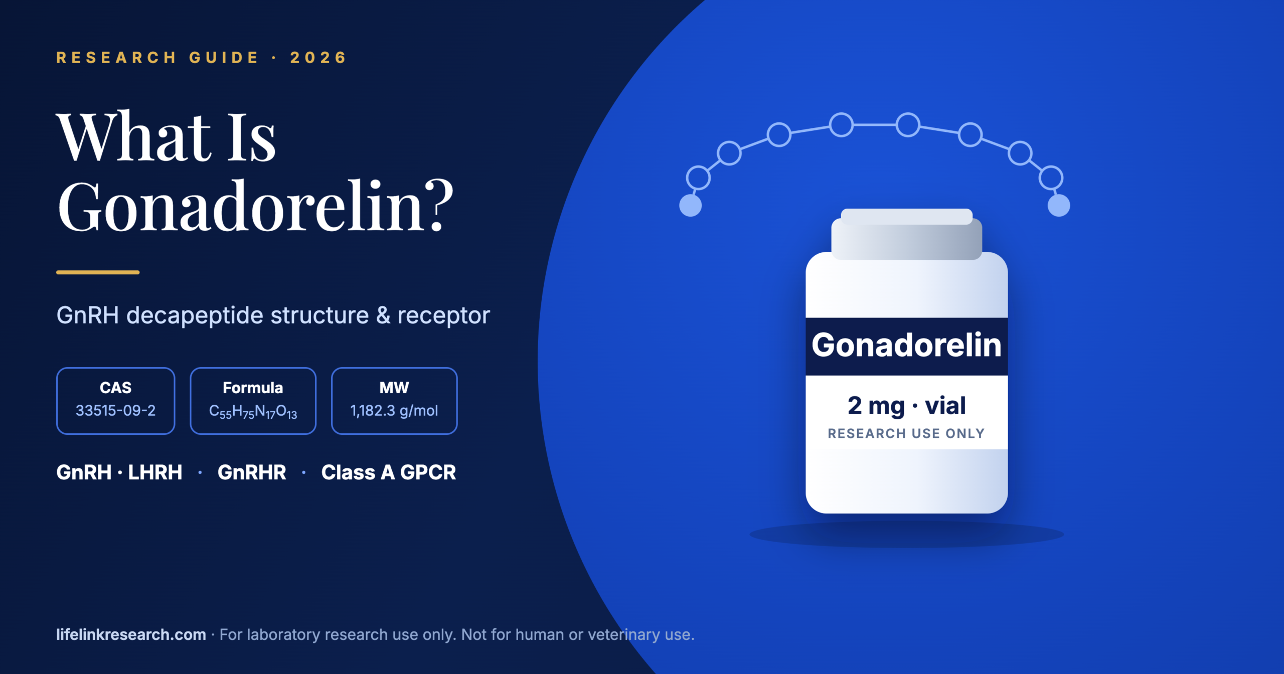
Gonadorelin is the synthetic decapeptide carrying the same sequence as mammalian GnRH-I, the peptide that gave its receptor a name. Ten residues, both ends chemically blocked, and a single G protein-coupled receptor to bind: on paper it looks like one of the simplest peptides a laboratory can work with.
The pharmacology underneath is less simple. This overview covers the sequence and structure, the salt forms that change what a vial actually contains, the GnRH receptor and its unusual architecture, the signalling readouts used to measure it, how to verify a batch, and where the compound stands with the FDA. New to peptide reagents? Start with our introduction to research peptides.
| Fact | Value |
| Name | Gonadorelin |
| Synonyms | GnRH, LHRH (GnRH-I) |
| Sequence | pGlu-His-Trp-Ser-Tyr-Gly-Leu-Arg-Pro-Gly-NH₂ |
| Length | 10 residues |
| Molecular weight | 1,182.3 g/mol, free base (PubChem CID 638793) |
| CAS, free base | 33515-09-2 |
| CAS, acetate | 34973-08-5 (diacetate tetrahydrate: 52699-48-6) |
| Common salt | Acetate |
| Target | GnRH receptor (GnRHR, gene GNRHR), class A GPCR |
| Primary coupling | Gq/11 |
| Form | Lyophilized powder |
| Status | Research use only |
What Is Gonadorelin?
Gonadorelin is the synthetic form of gonadotropin-releasing hormone (GnRH-I), a 10-amino-acid peptide with a blocked pyroglutamate N-terminus and an amidated C-terminus. It binds the GnRH receptor (GnRHR), a class A G protein-coupled receptor, and serves in research as the reference agonist for that receptor.
The name belongs to the manufactured material. “Gonadorelin” is the international nonproprietary name (INN) assigned to the synthetic peptide, which is why catalogues, pharmacopoeias and regulatory records use it while the physiology literature says GnRH.
Decapeptides of this family turn up across vertebrates with small sequence variations, and the mammalian form is the one gonadorelin reproduces. For a laboratory, the appeal is that a short synthetic sequence gives you the same ligand the receptor evolved around, with none of the batch variability of extracted material.
Gonadorelin vs GnRH: What’s the Difference?
Chemically there is none: gonadorelin has the same 10-residue sequence as native mammalian GnRH-I. The difference is origin and form. “GnRH” names the endogenous peptide, while “gonadorelin” names the synthetic material, usually supplied as an acetate salt, so its batch COA reports salt and net peptide content.
So the two words point at the same molecule from different angles. One is a biological entity, the other is a manufactured reagent with a lot number.
LHRH, short for luteinising hormone-releasing hormone, is an older synonym for the same peptide and still appears in literature from the 1970s and 1980s, along with catalogue listings that have never been renamed.
A second human decapeptide, GnRH-II, exists and differs at three positions. Its mature sequence reads pGlu-His-Trp-Ser-His-Gly-Trp-Tyr-Pro-Gly-NH₂ (UniProt O43555), so positions 5, 7 and 8 carry histidine, tryptophan and tyrosine where GnRH-I carries tyrosine, leucine and arginine. A type II receptor exists in many vertebrates, though in humans the gene carries a frameshift and is not expressed as a full receptor.
What Is the Structure of Gonadorelin?
Gonadorelin is a linear decapeptide: pGlu-His-Trp-Ser-Tyr-Gly-Leu-Arg-Pro-Gly-NH₂. Both ends are blocked, by a pyroglutamate ring at the N-terminus and an amide at the C-terminus. Glycine at position 6 allows a β-turn that folds the peptide into its receptor-binding shape, and positions 6 and 10 are where most synthetic analogs are modified.
The two blocked termini are the first thing to notice. Neither end carries the free amine or free acid a standard synthetic peptide would, which changes both its chemistry and the mass you should expect on a certificate.
Residue by residue
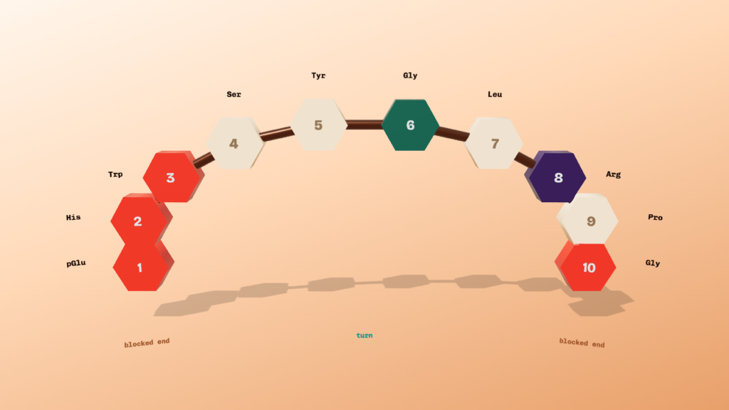
Gonadorelin decapeptide showing all ten residues from pGlu to Gly-amide, with both blocked ends and the glycine 6 turn highlighted
| Position | Residue | Structural note |
| 1 | pGlu | Cyclised glutamine, blocks the N-terminus |
| 2 | His | Part of the agonist-activity region |
| 3 | Trp | Part of the agonist-activity region |
| 4 | Ser | Flexible linker toward the turn |
| 5 | Tyr | Proposed receptor contact |
| 6 | Gly | Permits the β-turn |
| 7 | Leu | Hydrophobic, follows the turn |
| 8 | Arg | Required for high-affinity binding |
| 9 | Pro | Rigid, sets the C-terminal geometry |
| 10 | Gly-NH₂ | Amidated, blocks the C-terminus |
The Gly6 β-turn
Flanagan and Manilall (2017) describe the predominant conformer as “a β-turn that brings the amino- and carboxy-termini close together,” so the peptide folds back on itself. Their review assigns distinct jobs to the two ends: the N-terminal residues pGlu1, His2 and Trp3 drive agonist activity, while the C-terminal residues, “particularly Arg8, are necessary for high affinity binding.”
Why the native sequence is short-lived in assay matrices
Glycine at position 6 occupies the middle of the molecule with no side chain, and the Tyr5-Gly6 and Gly6-Leu7 bonds around it are the classic cleavage points for peptidases present in serum-containing media and tissue homogenates. Plan assay timing around that, and run a vehicle-matched control, since the peptide’s working life in a buffer differs from its life in a complex matrix.
How analogs change positions 6 and 10
Two chemical strategies dominate the analog literature, and both target the turn or the tail. Substituting a D-amino acid at position 6 stabilises the β-turn, a change that Flanagan and Manilall note increases receptor-binding affinity. Replacing the C-terminal Gly10-NH₂ with an ethylamide group is the other common modification. Both changes also remove a preferred cleavage site, which is why analog chemistry and stability chemistry tend to arrive together.
What Is the GnRH Receptor (GnRHR)?
The GnRH receptor (GnRHR, gene GNRHR) is a class A, seven-transmembrane G protein-coupled receptor. The mammalian type I receptor is unusual because it lacks the C-terminal tail most GPCRs carry, which is linked to its distinctive desensitisation behaviour in cell studies. It is studied in gonadotroph-derived cell lines such as αT3-1 and LβT2.
Receptor architecture
GnRHR belongs to class A, the rhodopsin-like family, and its fold is the standard “bundle of seven membrane-spanning α-helical segments.” Mutagenesis work has mapped several contacts between receptor and ligand: Asp7.32(302) is proposed to form a salt bridge with Arg8 of the peptide, and Tyr6.58(290) has been proposed as a contact for Tyr5. Other assignments remain open, and Flanagan and Manilall call it “uncertain whether Lys3.32(121) directly contacts GnRH or initiates receptor activation.”
The missing C-terminal tail
Here is the structural oddity. The mammalian type I receptor is “characterized by absence of a cytoplasmic carboxy-terminal tail,” the segment where most GPCRs are phosphorylated and where arrestin is recruited. Flanagan and Manilall report that the receptor “does not activate β-arrestin-dependent signaling,” leaving its output to run through G protein coupling. Type II receptors in other species do carry the tail, which makes the pair a natural comparison for studying what the tail contributes. Receptors in the same broad GPCR superfamily vary widely in this respect, and a useful contrast is B7-33 and the RXFP1 receptor, where a large extracellular domain does work that GnRHR handles within the helical bundle.
Cell models
Three systems carry most of the published work. The αT3-1 and LβT2 lines are gonadotroph-derived and express the receptor natively, with LβT2 the more mature of the two. HEK293 cells transfected with GnRHR give a cleaner background for binding and mutagenesis studies, which is where most receptor residue mapping has been done.
How Does Gonadorelin Signal Through the GnRH Receptor?
Gonadorelin binding activates GnRHR coupling to Gq/11, which stimulates phospholipase C-β. That produces IP3 and diacylglycerol, raising intracellular calcium and activating protein kinase C, followed by MAPK/ERK phosphorylation. In the laboratory these steps are measured as IP accumulation, calcium flux and pERK readouts.
The cascade runs in a fixed order, so each readout reports on a different depth of the pathway. Choosing between them is mostly a question of how far downstream the question lies and how fast the signal moves. Timing is the part that catches people out: a calcium response rises and falls within seconds to a couple of minutes, pERK peaks over several minutes, and IP accumulation is read as a total built up across half an hour or more. Sample at the wrong moment and a real response reads as no response at all.
| Readout | What it measures | Typical format |
| Radioligand binding | Affinity of a ligand for the receptor | Competition binding on membranes or whole cells |
| IP-One / IP accumulation | PLC-β activity, one step after Gq/11 | HTRF or similar plate assay, accumulation over 30 to 60 minutes |
| Ca²⁺ flux | Release of intracellular calcium stores | Dye-loaded cells read on a plate reader, seconds to minutes |
| pERK1/2 | MAPK activation further downstream | Western blot or plate-based phospho-assay, minutes |
| β-arrestin recruitment | Arrestin engagement at the receptor | Reporter assay; expect little signal at the tail-less type I receptor |
That last row is the practical consequence of the missing C-terminal tail. A β-arrestin assay that gives a flat response at GnRHR is reporting receptor biology, so confirm the assay with a positive-control receptor before concluding anything about the ligand.
Why Does Pulse Frequency Count in GnRH Receptor Research?
In cell and perifusion studies, continuous exposure to gonadorelin leads to receptor desensitisation and uncoupling, while intermittent pulses keep receptor signalling responsive. That is why pulse frequency is a key variable in GnRHR research design, and why perifusion systems that deliver timed pulses are used alongside static cell assays.
A static well of cells bathed in agonist for an hour and a perifusion chamber receiving five-minute pulses are two different experiments, and they can return different answers from the same compound.
Tsutsumi and colleagues (Journal of Biological Chemistry, 2010) examined signalling responses to pulsatile GnRH in LβT2 cells and showed that pathway activity tracks the pulse pattern as much as the total amount delivered. For anyone designing receptor work, three variables follow from that: pulse interval, pulse duration and washout between pulses, all of which belong in the methods section alongside concentration.
How Is Gonadorelin Used in Research?
Gonadorelin is used as the reference agonist in GnRH receptor studies: as the standard in binding and signalling assays, as the parent sequence for structure-activity work on analogs, and to study receptor desensitisation and internalisation in cell lines. It is supplied for research use only and not for any medical use.
| Source | Year | Study type | Assay readout |
| Flanagan CA, Manilall A. Front Endocrinol 8:274 | 2017 | Review of receptor structure and ligand binding | Binding determinants, receptor-residue contacts |
| Tsutsumi R et al. J Biol Chem 285:20262 | 2010 | In vitro, LβT2 gonadotroph cell line | Signalling responses under pulsatile delivery |
| Manilall A et al. Mol Cell Endocrinol 481:53 | 2019 | In vitro, receptor mutants in transfected cells | Ligand affinity and receptor activation |
What Is Gonadorelin Acetate?
Gonadorelin acetate is the peptide supplied as an acetate salt, the most common form for research material. The salt and any bound water add mass, so net peptide content is lower than the vial weight. Other forms, such as the diacetate tetrahydrate, exist, and the batch COA should state which salt is present.
The arithmetic has consequences at the bench. Weigh out 2 mg of acetate salt, treat it as 2 mg of peptide, and every concentration in the experiment drifts high by whatever fraction the counterion and water contribute. Certificates that report a measured peptide content let you correct for it, which is covered in our guide to net peptide content on a COA.
The scale of the correction is easy to pin down. PubChem lists the monoacetate at 1,242.3 g/mol against 1,182.3 for the free base, so the counterion alone accounts for about 5 percent of the weight, and the tetrahydrate form carries bound water on top of that. A 2 mg vial weighed as peptide is therefore closer to 1.9 mg of peptide before any residual solvent or water is counted.
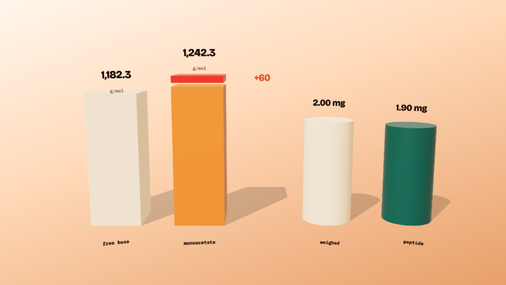
Comparison of gonadorelin free base at 1,182.3 g/mol against the monoacetate at 1,242.3 g/mol, and a 2.00 mg weighed vial against about 1.90 mg of peptide
How Do You Verify Gonadorelin Quality?
Confirm identity by mass spectrometry, where the observed mass should match roughly 1,182 Da for the free base, and purity by HPLC, then compare both with the batch result. Check that the COA names the salt form and reports net peptide content. Store the lyophilized powder cold, dry and away from light, as the label states.
Identity by mass spectrometry
The expected average mass is 1,182.3 Da for the free base, formula C55H75N17O13. Remember that the blocked termini are part of that figure: a report landing about 17 Da high suggests an unconverted glutamine at position 1, and about 1 Da high suggests a free acid where the C-terminal amide should be. See HPLC vs mass spectrometry for how the two methods divide the work.
Purity by HPLC
Read purity by HPLC, see the batch result, for the specific lot in hand. For what the figures mean in practice, see what 98% vs 99% purity means.
Third-party versus in-house testing
Who ran the assay carries as much weight as the number it produced. Our guide to third-party vs in-house testing sets out what each arrangement does and does not tell you.
Storage
Keep the sealed, lyophilized vial cold, dry and shielded from light, following the condition printed on the label. Gonadorelin is soluble in water for assay preparation. Our peptide storage guide covers handling of the dry form.
What Is Gonadorelin’s Regulatory Status in the US?
Gonadorelin has a history as an FDA-approved human drug product and as the active ingredient in approved animal drug products, which sets it apart from most research peptides. As of October 2026, research-grade gonadorelin is sold for laboratory use only. Check FDA sources for current product and compounding status.
| Date | Event | Primary source |
| 10 March 1978 | Cystorelin (gonadorelin diacetate tetrahydrate) approved under NADA 098-379 as an animal drug product | Animal Drugs @ FDA |
| 30 September 1982 | Factrel (gonadorelin hydrochloride) approved under NDA 018123; marketing status now listed as discontinued | Drugs@FDA |
| 10 October 1989 | Lutrepulse Kit (gonadorelin acetate) approved under NDA 019687; marketing status now listed as discontinued | Drugs@FDA |
| October 2026 | Both human products remain listed as discontinued in Drugs@FDA. Research-grade material is sold for laboratory use only | Drugs@FDA, read 12 October 2026 |
That approval history is the detail to carry away. Most research peptides have never held a US drug approval, so their status question is simple. Gonadorelin’s is a layered one: approved products that are no longer marketed, separate animal drug approvals that remain active, and research-grade material sold under a different framework entirely. Compounding categories are a further separate question, and our explainer on how FDA compounding lists work sets out how those lists operate.
Gonadorelin FAQs
Is gonadorelin the same as LHRH?
Yes, as a molecule. LHRH is an older name for the same decapeptide, used widely before GnRH became standard. Catalogues and papers from the 1970s and 1980s often use LHRH, so a literature search should cover both terms plus gonadorelin.
Is gonadorelin a peptide or a hormone?
Gonadorelin is a synthetic peptide with the same sequence as a peptide hormone. The word “hormone” describes the role the endogenous molecule plays, while “gonadorelin” describes manufactured material supplied as a laboratory reagent with a batch number and a certificate.
What kinds of compounds act at the GnRH receptor?
Two broad classes. Agonist analogs typically carry a D-amino acid at position 6 and often a modified C-terminus, which raises binding affinity and resists cleavage. Antagonist analogs carry several substitutions across the N-terminal residues that abolish the agonist activity while keeping receptor binding.
What is the molecular weight of gonadorelin?
1,182.3 g/mol for the free base, formula C55H75N17O13 (PubChem CID 638793). Material supplied as an acetate salt weighs more per vial, so check the salt form and the net peptide content on the certificate before calculating a concentration.
Does Life Link Research supply gonadorelin with a batch COA?
Yes. Our gonadorelin 2 mg research peptide ships with a batch-matched certificate from a third-party laboratory, covering identity by mass spectrometry and purity by HPLC for that lot.
References
| Source | Date | Type |
| Flanagan CA, Manilall A. Gonadotropin-releasing hormone (GnRH) receptor structure and GnRH binding. Front Endocrinol 8:274. https://pmc.ncbi.nlm.nih.gov/articles/PMC5662886/ | 2017 | Review |
| Tsutsumi R et al. Signaling responses to pulsatile gonadotropin-releasing hormone in LβT2 gonadotrope cells. J Biol Chem 285:20262. https://pmc.ncbi.nlm.nih.gov/articles/PMC2888439/ | 2010 | Primary research |
| Manilall A et al. Glu2.53(90) of the GnRH receptor is part of the conserved G protein-coupled receptor structure. Mol Cell Endocrinol 481:53. https://pubmed.ncbi.nlm.nih.gov/30476558/ | 2019 | Primary research |
| PubChem, gonadorelin, CID 638793. https://pubchem.ncbi.nlm.nih.gov/compound/638793 | Accessed 12 Oct 2026 | Chemical database |
| UniProt O43555, progonadoliberin-2. https://www.uniprot.org/uniprotkb/O43555/entry | Accessed 12 Oct 2026 | Sequence database |
| Drugs@FDA, NDA 018123 and NDA 019687. https://www.accessdata.fda.gov/scripts/cder/daf/index.cfm?event=overview.process&ApplNo=018123 | Accessed 12 Oct 2026 | Regulatory record |
| Animal Drugs @ FDA, NADA 098-379. https://animaldrugsatfda.fda.gov/adafda/views/ | Accessed 12 Oct 2026 | Regulatory record |
For Research Use Only. Not for use in diagnostic or therapeutic procedures. See our terms and research-use policy.
What Is B7-33? Relaxin-2 Analog Research Overview (2026)
Quick Answer B7-33 is a synthetic single-chain peptide built from part of the B chain of human relaxin-2, and it binds the…
Read article
Peptide Blends vs Single Vials: How to Choose for Research (2026)
For Research Use Only. Not for use in diagnostic or therapeutic procedures. Choosing between peptide blends vs single peptides comes down to…
Read article
What Is SLU-PP-332? Structure, Research Use & US Supply
Quick Answer SLU-PP-332 is a synthetic small-molecule agonist of the estrogen-related receptors ERRŒ±, ERRŒ≤ and ERRŒ≥ (CAS 303760-60-3, PubChem CID 5338394). It…
Read articleResearch-grade peptides
99%+ purity. Verified every batch.
Identity and purity tested on every batch by third-party labs, Janoshik Analytical and US-based laboratories. Shipped same day from California.
Shop all peptides