• Order within –h –m
  • Free shipping on orders over $250
  • Free Bacteriostatic Water 30ml with $500+ purchase
  • Every batch tested by an independent lab
  • For laboratory and research use only. Not for human consumption

Category: Comparison

  • Selank vs Semax: Two Pro-Gly-Pro Peptides Compared by Structure and Identity

    Selank vs Semax: Two Pro-Gly-Pro Peptides Compared by Structure and Identity

    Quick Answer

    Selank and Semax are both seven-residue synthetic peptides that end in the same Pro-Gly-Pro tail, but their first four residues come from different parent peptides. Selank (Thr-Lys-Pro-Arg-Pro-Gly-Pro) extends tuftsin and weighs 751.9 g/mol; Semax (Met-Glu-His-Phe-Pro-Gly-Pro) extends ACTH(4-7), weighs 813.9 g/mol and contains sulfur.

    Key takeaways

    • Selank: tuftsin + PGP, C33H57N11O9, 751.9 g/mol, CAS 129954-34-3 (also TP-7)
    • Semax: ACTH(4-7) + PGP, C37H51N9O10S, 813.9 g/mol, CAS 80714-61-0
    • The shared Pro-Gly-Pro tail slows breakdown by peptidases
    • CAS 4037-01-8, often listed beside Semax, belongs to ACTH(4-10)
    • By mass spectrometry, the two are 62 Da apart on a COA

    Selank is tuftsin with three extra amino acids. Semax is a fragment of ACTH with the same three extras. Both are seven-residue synthetic peptides, and both end in an identical tail: proline, glycine, proline.

    That shared ending is why Selank and Semax are such a common pairing, and it hides two quite different molecules. Their front halves come from unrelated parent peptides, their weights differ by 62 Da, and only one of them contains sulfur.

    This guide compares Selank vs Semax by structure and identity: sequence, origin, formula, molecular weight, CAS number, the salt and N-acetyl forms that change those numbers, and what each COA should show. Our Semax + Selank research kit supplies both, and it’s the example used throughout.

    What’s the Difference Between Selank and Semax?

    Quick answer: Selank and Semax are both seven-amino-acid synthetic peptides ending in Pro-Gly-Pro, but they come from different parent sequences. Selank extends tuftsin (Thr-Lys-Pro-Arg), while Semax extends the ACTH(4-7) fragment (Met-Glu-His-Phe). They differ in formula, molecular weight (751.9 vs 813.9 g/mol) and CAS number, and only Semax contains sulfur.

    PropertySelankSemax
    Parent peptideTuftsin (Thr-Lys-Pro-Arg)ACTH(4-7) (Met-Glu-His-Phe)
    Full sequenceThr-Lys-Pro-Arg-Pro-Gly-ProMet-Glu-His-Phe-Pro-Gly-Pro
    Length7 residues7 residues
    FormulaC33H57N11O9C37H51N9O10S
    Molecular weight751.9 g/mol813.9 g/mol
    CAS129954-34-380714-61-0
    SulfurNoYes, from methionine
    Other namesTP-7ACTH(4-7)PGP
    OriginInstitute of Molecular Genetics, Russian Academy of SciencesInstitute of Molecular Genetics, Russian Academy of Sciences
    PubChem CID117656009811102

    Read the table from the bottom up, and the pattern is clear. Same institute, same tail, same length, different starting material.

    What Is Selank?

    Quick answer: Selank (also called TP-7) is a synthetic heptapeptide, Thr-Lys-Pro-Arg-Pro-Gly-Pro. It consists of tuftsin, a natural tetrapeptide, with a Pro-Gly-Pro extension added at the C-terminus. It was developed at the Institute of Molecular Genetics of the Russian Academy of Sciences. Its formula is C33H57N11O9 and its CAS number is 129954-34-3.

    Sequence and the Tuftsin Core

    The Selank peptide is built in two parts. The first four residues are tuftsin, a naturally occurring tetrapeptide: threonine, lysine, proline, arginine. Two of those, lysine and arginine, carry basic side chains, so Selank is strongly positively charged at neutral pH. The added Pro-Gly-Pro makes residues 5 to 7, and the chain ends in a free carboxyl group. With three prolines in seven positions, Selank is a proline-rich, flexible peptide with no fixed secondary structure.

    Formula, MW, CAS

    PubChem CID 11765600 lists Selank as C33H57N11O9, with a molecular weight of 751.9 g/mol and a monoisotopic mass of 751.43 Da. The record carries CAS 129954-34-3. Tuftsin alone, by comparison, is C21H40N8O6 at 500.6 g/mol (CID 156080).

    Assay Targets Reported in the Literature

    Published work on Selank centres on GABA receptor systems, measured in two ways:

    • Rodent gene-expression study: Volkova et al., 2016 used real-time PCR in rats to measure expression of genes linked to GABA signalling, comparing Selank with GABA itself.
    • Rodent receptor-binding study: Vasil’eva et al., 2016 measured radioligand binding at GABA and NMDA receptor sites in two mouse strains.

    Both are animal studies, and each result belongs to its own model. Single-compound work uses our Selank 10 mg vial.

    What Is Semax?

    Quick answer: Semax is a synthetic heptapeptide, Met-Glu-His-Phe-Pro-Gly-Pro. It keeps the ACTH(4-7) fragment and replaces residues 8 to 10 with a Pro-Gly-Pro extension. It comes from the same Institute of Molecular Genetics team in Moscow, and PubChem lists it as C37H51N9O10S under CAS 80714-61-0.

    Sequence: Why “ACTH(4-7) + PGP” and Not ACTH(4-10)

    The Semax peptide is built the same way, from a different starting point. ACTH, adrenocorticotropic hormone, is a 39-residue peptide. Its fragment 4-10 reads Met-Glu-His-Phe-Arg-Trp-Gly. Semax keeps the first four of those residues and swaps the last three, Arg-Trp-Gly, for Pro-Gly-Pro. So Semax is accurately described as ACTH(4-7)PGP. Calling it “an ACTH(4-10) analog” is common, and it’s where a lot of the identity confusion starts.

    Formula, MW, CAS

    PubChem CID 9811102 lists Semax as C37H51N9O10S, 813.9 g/mol, with a monoisotopic mass of 813.35 Da and CAS 80714-61-0. A second number, CAS 4037-01-8, often appears beside Semax online. PubChem assigns that CAS to ACTH(4-10) itself, C44H59N13O10S at 962.1 g/mol (CID 123787), so it describes the parent fragment.

    Assay Targets Reported in the Literature

    Semax studies lean heavily on transcription profiling:

    • Rodent gene-expression study: Medvedeva et al., 2014 ran a genome-wide transcriptional analysis in a rat model, mapping which genes changed expression after Semax.
    • Rodent receptor-gene study: Liu et al., 2025 examined the μ-opioid receptor gene Oprm1 in mice, including deubiquitination assays.

    As with Selank, both are animal studies and describe their own models only. Single-compound work uses our Semax 10 mg vial.

    Why Do Selank and Semax Both End in Pro-Gly-Pro?

    Quick answer: The Pro-Gly-Pro (PGP) tail was added to both peptides to slow breakdown by peptidases. Short natural fragments like tuftsin and ACTH(4-7) are cleaved quickly in biological matrices, while proline-rich sequences resist many exopeptidases. Stability studies with purified peptidases and rat plasma compared proline-containing peptides and their breakdown products.

    Proline is the key. Its side chain loops back onto its own backbone nitrogen, which locks the peptide bond into an awkward shape for many enzymes. Exopeptidases that trim residues one at a time from either end tend to stall when they reach a proline. Placing Pro-Gly-Pro at the C-terminus guards the end that carboxypeptidases attack.

    The Institute of Molecular Genetics group behind both peptides has tested this directly. Shevchenko et al., 2019 reviewed in vitro hydrolysis of glyproline peptides by leucine aminopeptidase, dipeptidases, and carboxypeptidases B and Y, and in rat plasma. An earlier 2013 study from the same team tracked the cleavage of simple glyprolines by the same enzymes. Both describe a single design idea applied to two unrelated starting sequences.

    Sequence Comparison Table

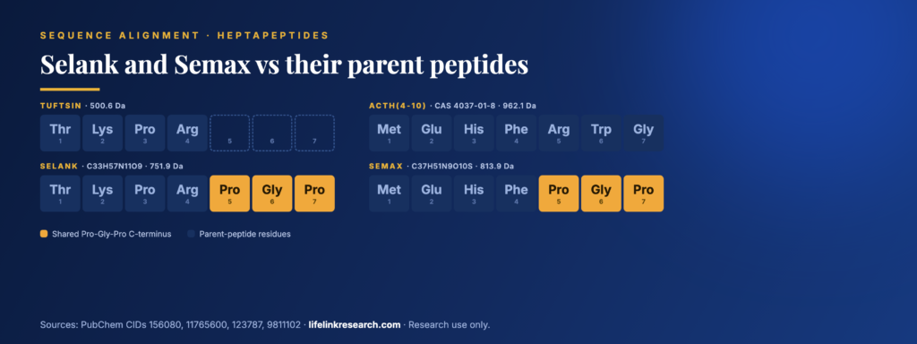
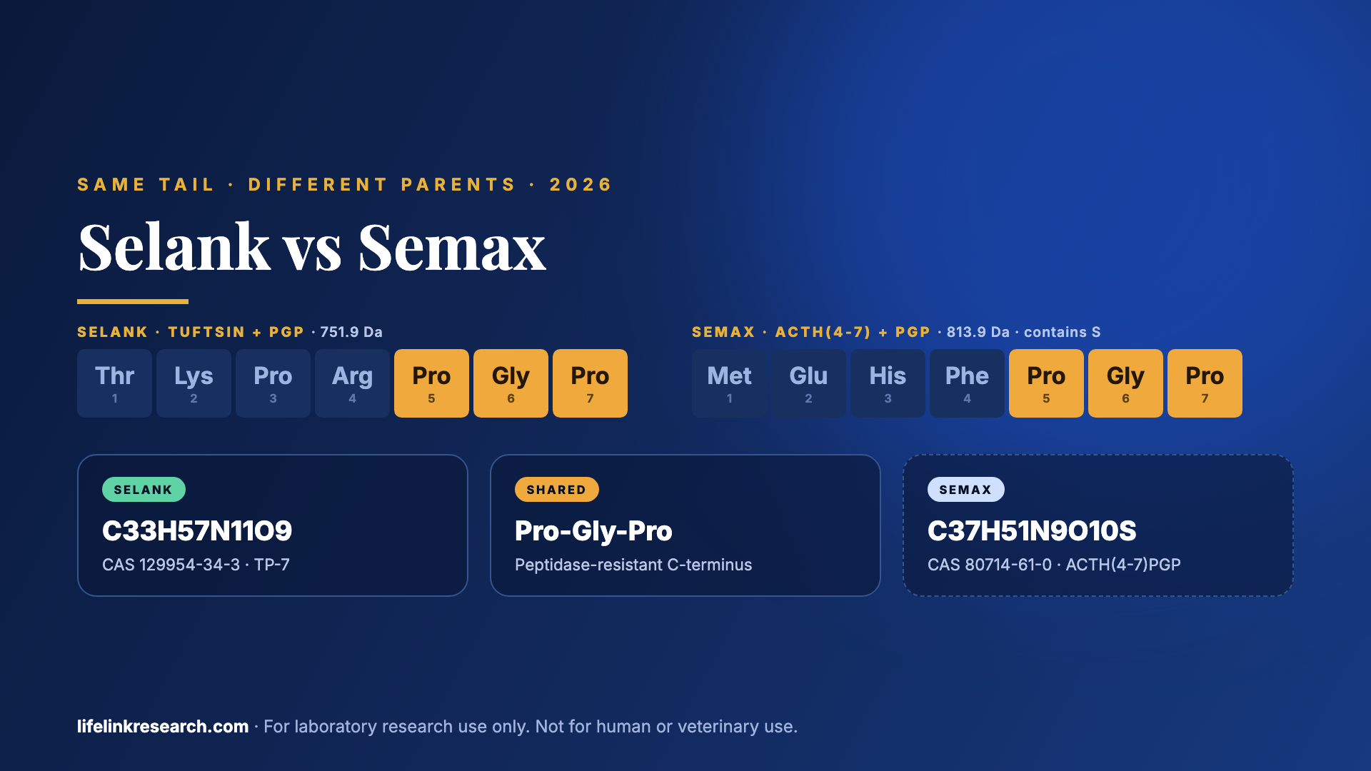
    Lined up by position, the four peptides show where Selank and Semax come from and where they meet:

    PositionTuftsinSelankACTH(4-10)Semax
    1ThrThrMetMet
    2LysLysGluGlu
    3ProProHisHis
    4ArgArgPhePhe
    5noneProArgPro
    6noneGlyTrpGly
    7noneProGlyPro
    MW (g/mol)500.6751.9962.1813.9
    Selank and Semax aligned with their parent peptides, with the shared Pro-Gly-Pro C-terminus highlighted.

    Selank and Semax aligned with their parent peptides, with the shared Pro-Gly-Pro C-terminus highlighted.

    Positions 5 to 7 are identical in Selank and Semax. Positions 1 to 4 share nothing, and that’s exactly the part a mass spectrometer reads as a 62 Da difference in weight. Glance down the MW row and the effect of the PGP swap is visible too: Semax is lighter than ACTH(4-10) because Pro-Gly-Pro weighs less than Arg-Trp-Gly.

    N-Acetyl and Salt Forms: Different Molecules, Different MW

    Quick answer: N-acetyl Semax amidate and N-acetyl Selank are chemically different molecules from standard Semax and Selank. Acetylation and C-terminal amidation change the formula and molecular weight. Salt forms, such as Selank diacetate, also raise the weight on a COA. Always check that the COA’s molecular weight matches the exact form named on the label.

    FormChangeFormulaMW (g/mol)PubChem CID
    SelankNoneC33H57N11O9751.911765600
    Selank acetateOne acetic acid as saltC35H61N11O11811.9155489759
    Selank diacetateTwo acetic acids as saltC37H65N11O13872.0155804769
    N-acetyl SelankAcetyl group on the N-terminusC35H59N11O10793.9133082488
    SemaxNoneC37H51N9O10S813.99811102
    N-acetyl Semax amidateN-terminal acetyl plus C-terminal amideC39H54N10O10S855.0172638603

    These two kinds of change show up differently on a COA. Salt forms change the weight of the powder, while the mass spectrometer still sees the bare peptide ion. Acetylation and amidation change the peptide itself, so they move the MS result too.

    How to Tell Selank and Semax Apart on a COA

    Quick answer: Mass spectrometry separates them clearly. Selank should show a mass near 751.9 Da and Semax near 813.9 Da, about 62 Da apart. Only Semax contains sulfur, from its methionine. Each vial in a kit needs its own COA with HPLC purity, an MS identity result matching that peptide, and a lot number matching the vial.

    Expected MS Masses ([M+H]⁺ and [M+2H]²⁺)

    Small peptides like these usually appear as singly and doubly protonated ions in electrospray MS. Calculated from the monoisotopic masses:

    IonSelank (751.43)Semax (813.35)
    [M+H]⁺m/z ≈ 752.4m/z ≈ 814.4
    [M+2H]²⁺m/z ≈ 376.7m/z ≈ 407.7

    On our current Semax report from Krause Analytical, issued 13 July 2026, the spectrum shows its two main ions at m/z 815.1 and 408.4, with the lab recording a mass spectral match as “ID Confirmed.” Lower-resolution instruments often read the most abundant isotope peak, which reads a little above the monoisotopic value. Two peptides this close in size need identity confirmed by mass spectrometry, not purity alone.
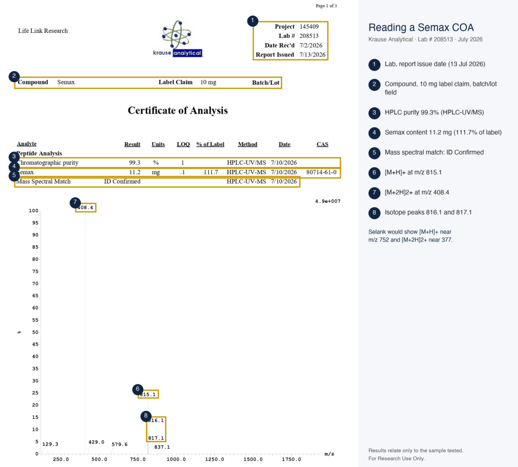
    Semax COA from Krause Analytical (2026), with the purity, content, and mass-spec results labelled.

    Semax COA from Krause Analytical (2026), with the purity, content, and mass-spec results labelled.

    HPLC Purity and the Salt-Form Note

    The same Semax report gives chromatographic purity of 99.3% by HPLC-UV/MS, plus a measured content of 11.2 mg against a 10 mg label claim. Our current Selank report, from Prime Analytical Laboratories in March 2026, gives 99.21% HPLC purity. If a label names a salt form, its stated weight includes the counter-ion, so the peptide content per mg runs lower.

    Lot, Date and Lab Checks

    A Selank COA or Semax COA should carry a lot number that matches its vial, a test date that fits the batch, and a lab you can contact. In a two-vial kit, check each report against its own vial. Our guide on how to verify a peptide COA walks through all six checks.

    Why Are Selank and Semax Sold as a Kit?

    The kit format is about supply. The Balance kit holds two separate vials, 10 mg of Semax and 10 mg of Selank, and each vial keeps its own batch COA. Nothing is premixed, so each peptide can be weighed, tested, and used in a study on its own terms.

    The pairing makes sense for comparison work. Two heptapeptides with the same C-terminal design and different N-terminal sequences make a tidy matched set for studies that look at how the parent sequence shapes a result. Premixed vials and separate ones trade off convenience against control over the ratio.

    Are Selank and Semax FDA Approved?

    Quick answer: No. As of 31 October 2026, neither Selank nor Semax is an FDA-approved drug in the US; both are sold for laboratory research use only. Both were developed in Russia, where they hold national registrations as medicines.

    • FDA: the FDA’s bulk drug substances page, current as of 22 April 2026, lists “Selank acetate (TP-7)” and “Semax (heptapeptide)” among nominations that were withdrawn.
    • Research supply: both peptides are sold in the US as research chemicals for in-vitro laboratory work.

    FAQs

    Is Selank the same as Semax?

    No. Both are seven-residue peptides ending in Pro-Gly-Pro, but their first four residues differ completely. Selank starts with tuftsin (Thr-Lys-Pro-Arg); Semax starts with ACTH(4-7) (Met-Glu-His-Phe). They have different formulas, weights, and CAS numbers, and only Semax contains sulfur.

    What is the molecular weight of Selank and Semax?

    Selank weighs 751.9 g/mol (C33H57N11O9, CAS 129954-34-3). Semax weighs 813.9 g/mol (C37H51N9O10S, CAS 80714-61-0). The 62 Da difference is easy to see by mass spectrometry. A salt label changes the numbers too: the diacetate form of Selank comes to 872.0 g/mol.

    What does Pro-Gly-Pro do in Selank and Semax?

    Pro-Gly-Pro is a C-terminal extension that slows breakdown by peptidases. Proline’s ring structure makes the neighbouring peptide bonds hard for many exopeptidases to cut, so the tail shields the peptide’s C-terminal end from trimming enzymes. The design was applied to both parent sequences by the same institute.

    Is N-acetyl Semax the same as Semax?

    They’re related but distinct. N-acetyl Semax amidate carries an acetyl group on the N-terminus and an amide at the C-terminus. That changes the formula to C39H54N10O10S and the weight to 855.0 g/mol, about 41 Da more than Semax, so it shows a different mass on a COA.

    How do I verify a Selank or Semax COA?

    Check that the MS result matches the peptide: near 752 for Selank’s [M+H]⁺ and 814 for Semax’s. Then confirm HPLC purity, match the lot number to the vial, and contact the named lab to verify the report. In a kit, check both vials separately.

    What is in the Life Link Balance kit?

    The kit holds one 10 mg vial of Semax and one 10 mg vial of Selank, packed as two separate vials in one box. Nothing is premixed. Each vial has its own batch COA, and earlier reports for each product stay on its certificate page.

    Buy the Semax + Selank Kit With Batch COAs

    Our kit pairs one vial of each peptide at 10 mg, each with its own batch report. Identity and purity are tested on every batch through independent labs. To find a batch COA, open the single-peptide product, click View report, and match the batch number to the vial label. Orders placed before 2 PM PST ship the same day from California by UPS 2-Day Air.

    For Research Use Only. Not for use in diagnostic or therapeutic procedures.

    All products are supplied for in-vitro laboratory research by qualified professionals. Read the full research use disclaimer.

    References

  • CJC-1295 With DAC vs Without DAC (Mod GRF 1-29): Structure & Identity Compared

    CJC-1295 With DAC vs Without DAC (Mod GRF 1-29): Structure & Identity Compared

    Quick Answer

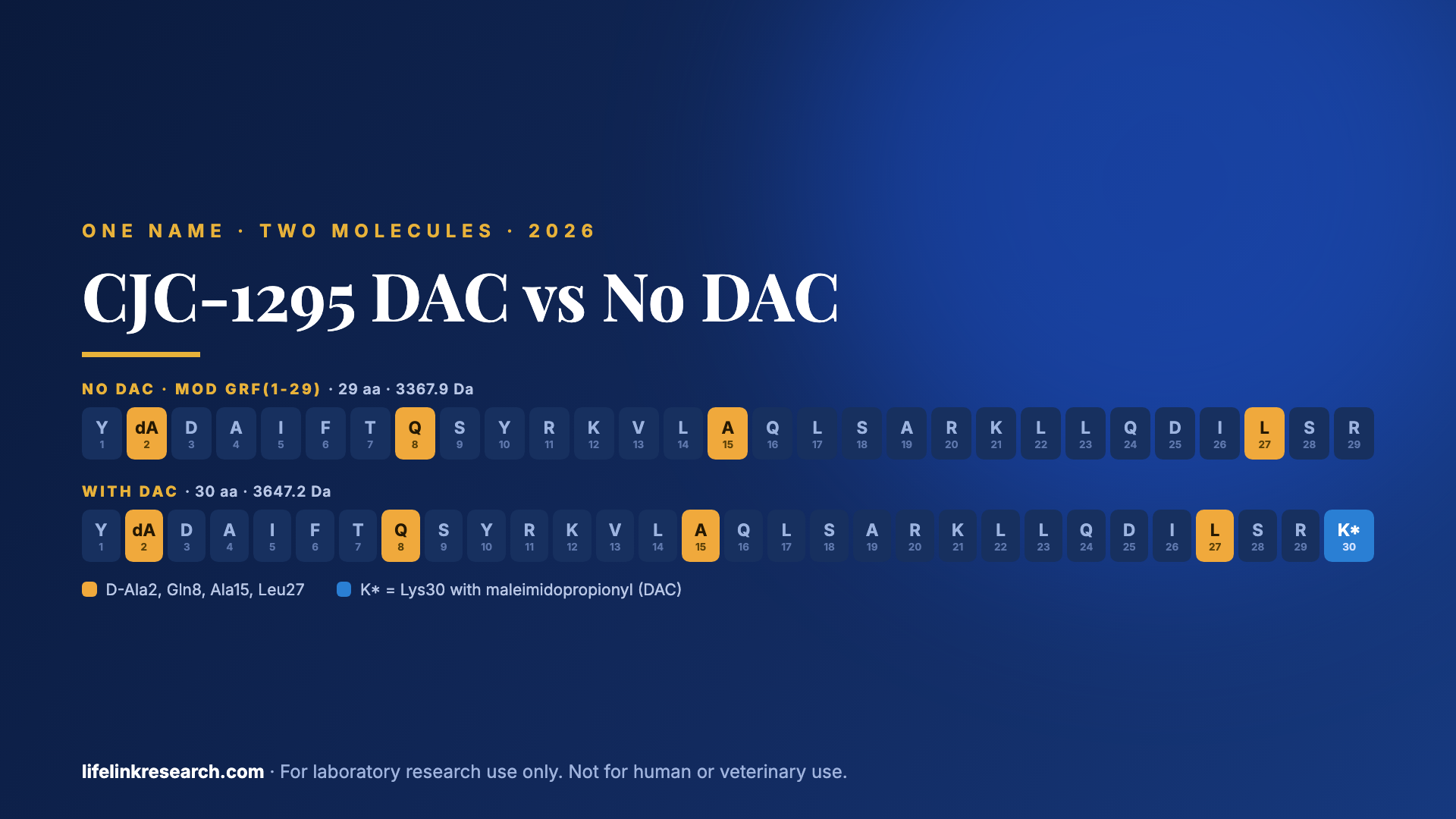
    CJC-1295 with DAC and CJC-1295 without DAC (Mod GRF 1-29) share the same 29-residue GHRH(1-29) core with four substitutions: D-Ala2, Gln8, Ala15 and Leu27. The DAC form adds a 30th residue, a lysine carrying a maleimidopropionyl group that bonds to albumin. That raises the weight from 3367.9 to 3647.2 g/mol, a difference mass spectrometry reads clearly.

    Key takeaways

    • Both forms share the tetrasubstituted GHRH(1-29) sequence: D-Ala2, Gln8, Ala15, Leu27
    • With DAC: 30 residues, C165H269N47O46, 3647.2 g/mol, CAS 446262-90-4
    • Without DAC (Mod GRF 1-29): 29 residues, C152H252N44O42, 3367.9 g/mol, CAS 863288-34-0
    • The DAC linker is a maleimidopropionyl group on Lys30 that reacts with albumin Cys34
    • Only a mass-spec result on the COA shows which form a vial holds

    CJC-1295 with DAC is a 30-residue GHRH analog carrying an albumin-reactive linker. CJC-1295 without DAC, better known as Mod GRF(1-29), is the same 29-residue core with no linker at all. One name, two molecules, separated by a single lysine and a small chemical hook.

    That difference is easy to state and surprisingly easy to lose on a label. In the CJC-1295 DAC vs no DAC comparison, the two forms differ by about 279 Da, carry different CAS numbers, and need different expected masses on a COA.

    This guide sets out exactly how they differ in sequence, structure, and identity data, and how to check which one a vial contains, using our CJC-1295 no DAC 5 mg as the worked example.

    What’s the Difference Between CJC-1295 With DAC and Without DAC?

    Quick answer: Both are synthetic analogs of GHRH(1-29) with the same four amino-acid substitutions. CJC-1295 with DAC adds a 30th residue, a lysine carrying a maleimidopropionyl group, which bonds covalently to albumin. CJC-1295 without DAC, also called Mod GRF(1-29), has no linker. They differ in formula, molecular weight (about 3647 vs 3368 Da), and CAS number.

    PropertyWith DACWithout DAC (Mod GRF 1-29)
    Length30 residues29 residues
    C-terminusLys30 amide, side chain modifiedArg29 amide
    Albumin linkerMaleimidopropionyl group on Lys30None
    FormulaC165H269N47O46C152H252N44O42
    Molecular weight3647.2 g/mol3367.9 g/mol
    CAS446262-90-4863288-34-0
    PubChem CID9197182056841945
    Name originConjuChem development codeSupplier name for the unlinked core
    ReceptorGHRH receptorGHRH receptor

    Everything above the linker row is identical chemistry. Everything below it follows from one extra residue.

    What Is CJC-1295 Without DAC (Mod GRF 1-29)?

    Quick answer: CJC-1295 without DAC is a 29-amino-acid analog of GHRH(1-29) with four substitutions (D-Ala2, Gln8, Ala15, Leu27) that make it more resistant to enzymatic breakdown in assays than the native sequence. It is also called Mod GRF(1-29) or tetrasubstituted GRF(1-29). It binds the GHRH receptor and carries no albumin-binding linker.

    Full Sequence

    Tyr-D-Ala-Asp-Ala-Ile-Phe-Thr-Gln-Ser-Tyr-Arg-Lys-Val-Leu-Ala-Gln-Leu-Ser-Ala-Arg-Lys-Leu-Leu-Gln-Asp-Ile-Leu-Ser-Arg-NH2

    In one-letter code, that’s Y(D-A)DAIFTQSYRKVLAQLSARKLLQDILSR-NH2. The C-terminal amide matches native GHRH(1-29)-NH2, and the D-alanine at position 2 is the only non-standard residue.

    Formula, MW, CAS

    PubChem CID 56841945 lists the 29-residue structure as C152H252N44O42, with a molecular weight of 3367.9 g/mol and a monoisotopic mass of 3365.89 Da. That record carries CAS 863288-34-0. No sulfur appears in the formula because the one methionine of the native sequence has been replaced.

    Why the Four Substitutions Make Chemical Sense

    Each change removes a known weak point in the native sequence:

    • D-Ala2: the enzyme DPP-IV clips GHRH between positions 2 and 3. A D-amino acid at position 2 resists that cut.
    • Gln8: native Asn8 can deamidate, slowly turning into aspartate or iso-aspartate in solution. Glutamine is far less prone to it.
    • Ala15: native Gly15 is flexible. Alanine favours the helix this region forms when it binds the receptor.
    • Leu27: native Met27 oxidises easily to methionine sulfoxide. Leucine has a similar shape and no sulfur to oxidise.

    What Is CJC-1295 With DAC?

    Quick answer: CJC-1295 with DAC is the same tetrasubstituted GHRH(1-29) sequence plus a C-terminal lysine whose side chain carries a maleimidopropionyl group, the “drug affinity complex”. In the original ConjuChem design, this maleimide forms a covalent bond with the free cysteine (Cys34) of serum albumin. It has 30 residues and a molecular weight of about 3647 Da.

    The DAC Linker: Maleimidopropionyl-Lysine

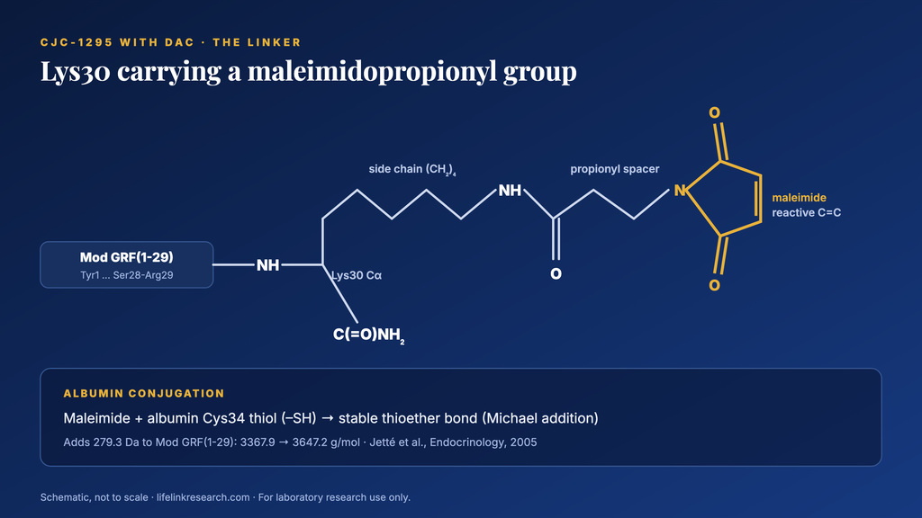
    The linker is small. A lysine is added as residue 30, and its side-chain amine (the ε-nitrogen) is acylated with 3-maleimidopropionic acid. That leaves a maleimide ring, a five-membered ring with a reactive carbon-carbon double bond, pointing away from the peptide. The C-terminus stays amidated.

    The DAC linker on Lys30: a maleimidopropionyl group attached to the lysine side chain.

    The DAC linker on Lys30: a maleimidopropionyl group attached to the lysine side chain.

    The maleimide does all the chemistry. The rest of the molecule is the same Mod GRF(1-29) core described above.

    Albumin Cys34 Conjugation

    Serum albumin carries one free thiol, on cysteine 34. Maleimides react with thiols in a Michael addition, forming a stable thioether bond. ConjuChem’s team built CJC-1295 around exactly that reaction, placing the maleimide where it could reach Cys34 without blocking the receptor-binding end, as Jetté and colleagues described in 2005. That paper combined ex vivo conjugation to human serum albumin with a rat study, and it is where the CJC-1295 name first appears in the literature.

    Formula, MW, CAS

    PubChem CID 91971820 lists CJC-1295 with DAC as C165H269N47O46, 3647.2 g/mol, with a monoisotopic mass of 3645.02 Da and CAS 446262-90-4.

    Sequence Comparison: Sermorelin vs Mod GRF 1-29 vs CJC-1295 DAC

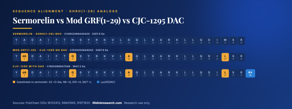
    All three share the same 29-residue backbone. Only five positions change across them:

    PositionSermorelin, GHRH(1-29)Mod GRF(1-29), no DACCJC-1295 with DAC
    1TyrTyrTyr
    2AlaD-AlaD-Ala
    3 to 7Asp-Ala-Ile-Phe-Thrsamesame
    8AsnGlnGln
    9 to 14Ser-Tyr-Arg-Lys-Val-Leusamesame
    15GlyAlaAla
    16 to 26Gln-Leu-Ser-Ala-Arg-Lys-Leu-Leu-Gln-Asp-Ilesamesame
    27MetLeuLeu
    28 to 29Ser-Argsamesame
    30nonenoneLys(maleimidopropionyl)
    C-terminusAmideAmideAmide
    Formula / MWC149H246N44O42S / 3357.9C152H252N44O42 / 3367.9C165H269N47O46 / 3647.2

    Sermorelin, Mod GRF(1-29) and CJC-1295 with DAC aligned residue by residue, with the changed positions highlighted.

    Sermorelin is the unmodified 1-29 sequence, with formula and weight from PubChem CID 16132413. The no-DAC form is therefore a separate molecule, about 10 Da heavier and sulfur-free. For a wider look at sermorelin, see ipamorelin vs sermorelin.

    Why Is “CJC-1295” Confusing? Naming Explained

    Quick answer: “CJC-1295” was originally the name of the DAC-linked molecule developed by ConjuChem. Suppliers later began selling the unlinked Mod GRF(1-29) as “CJC-1295 no DAC”, so one name now covers two compounds. Always confirm the form by molecular weight on the COA: about 3647 Da with DAC and about 3368 Da without.

    Name on a labelWhich moleculeExpected MW
    CJC-1295Usually with DAC, but check3647.2 or 3367.9
    CJC-1295 DACWith DAC3647.2
    CJC-1295 no DACWithout DAC3367.9
    Mod GRF(1-29)Without DAC3367.9
    Modified GRF 1-29Without DAC3367.9
    Tetrasubstituted GRF(1-29)Without DAC3367.9

    The confusion began when suppliers kept the familiar CJC-1295 name for the shorter, unlinked peptide. Even public databases trip over this. PubChem holds two records with the 29-residue formula, and both list “with DAC” and “without DAC” among their synonyms. The formula and weight are the reliable part of each record; the synonym lists are supplied by depositors.

    How Do Their Half-Lives Compare in Published Studies?

    Quick answer: Published figures differ by orders of magnitude. A human pharmacokinetic study of CJC-1295 with DAC (Teichman et al., 2006) reported an estimated half-life of 5.8 to 8.1 days, linked to albumin binding. Mod GRF(1-29) without DAC is usually described as having a half-life of about 30 minutes, a figure often stated without a primary source.

    The Teichman figure comes from two randomized, placebo-controlled trials in healthy adults, with the half-life estimated from plasma concentrations of the peptide. We couldn’t trace the 30-minute figure to a pharmacokinetic study of Mod GRF(1-29) itself, so treat it as an approximation. Ipamorelin’s numbers sit in a different range, which is where CJC-1295 and ipamorelin diverge.

    How to Tell Them Apart on a COA

    Quick answer: Mass spectrometry is the clearest check. CJC-1295 without DAC should show a mass near 3368 Da, and CJC-1295 with DAC near 3647 Da, about 279 Da higher because of the extra lysine and maleimidopropionyl group. HPLC purity alone can’t separate the two forms, so a COA must report an MS result that matches the label.

    Expected MS Masses and Charge States

    Electrospray ionisation adds several protons to a peptide this size, so the spectrum shows a ladder of multiply charged ions, one for each charge state. Dividing (mass + charge × 1.007) by the charge gives the expected positions:

    IonWithout DAC (3367.9)With DAC (3647.2)
    [M+3H]³⁺m/z ≈ 1123.6m/z ≈ 1216.7
    [M+4H]⁴⁺m/z ≈ 843.0m/z ≈ 912.8
    [M+5H]⁵⁺m/z ≈ 674.6m/z ≈ 730.4

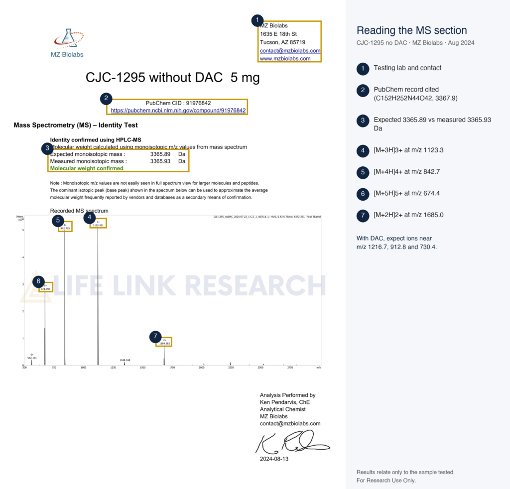
    Labs then convert the ladder back to one mass. On our earlier MZ Biolabs report pictured below, the no-DAC sample showed ions at m/z 1123.3, 842.7, and 674.4, and a measured monoisotopic mass of 3365.93 Da against 3365.89 expected. HPLC and mass spectrometry answer different questions about a DAC and no-DAC pair: how much is there, and which molecule it is.

    Mass-spec identity section from a Life Link Research CJC-1295 no DAC COA (MZ Biolabs), with each charge state labelled.

    Mass-spec identity section from a Life Link Research CJC-1295 no DAC COA (MZ Biolabs), with each charge state labelled.

    HPLC Purity and Retention

    HPLC gives purity as the main peak’s share of total area. Our current no-DAC report from Prime Analytical Laboratories, dated 5 May 2026, shows the main peak at 6.317 minutes with 99.30% of the area. The DAC form’s extra hydrophobic linker would shift its retention time, though only a side-by-side run on the same method can show by how much. That’s why purity and identity are separate lines on a good report: one says how clean the sample is, the other says what it is.

    Label, Lot and Lab Checks

    A CJC-1295 COA should name the form exactly, “no DAC” or “with DAC”, and the vial label should use the same wording. A report that just says “CJC-1295” leaves the main question open, so the MS result has to answer it. Then match the lot number, check the test date, and verify the report with the named lab. Our guide on how to verify a peptide COA covers all six checks.

    Receptor Target: GHRH-R

    Quick answer: Both forms bind the growth-hormone-releasing hormone receptor (GHRH-R), a class B G-protein-coupled receptor that signals through cAMP. In vitro, both are studied with receptor-binding and cAMP-accumulation assays. The DAC linker doesn’t change the receptor target; it changes how the molecule behaves in albumin-containing systems.

    A common set-up compares cells engineered to express human GHRH-R with the parent cell line that lacks it, so any cAMP signal can be tied to the receptor. In practice, assay buffers deserve attention too. A medium containing albumin can capture the DAC form through Cys34, while the no-DAC form stays free, so the two can behave differently in the same cell assay.

    CJC-1295 No DAC and Ipamorelin as a Blend

    The no-DAC form often appears in a 5/5 mg blend with ipamorelin, and the reason is receptor classes. CJC-1295 no DAC binds GHRH-R, a class B GPCR. Ipamorelin, a pentapeptide (Aib-His-D-2-Nal-D-Phe-Lys-NH2), binds the ghrelin receptor GHSR-1a, a class A GPCR. Two receptor families, one vial. Its COA therefore needs two identity results: the Mod GRF(1-29) mass near 3368 Da and the ipamorelin mass near 711.9 Da (C38H49N9O5, PubChem CID 9831659). Our CJC-1295 / Ipamorelin blend 5/5 mg holds 5 mg of each, and the guide to what is Ipamorelin covers its structure. The CJC-1295 + Ipamorelin blend guide covers that vial’s composition and COA.

    Is CJC-1295 FDA Approved?

    Quick answer: No. As of 31 October 2026, neither CJC-1295 with DAC nor Mod GRF(1-29) is an FDA-approved drug. Both are sold in the US for laboratory research use only.

    • Drug records: the FDA’s openFDA drug database returns no application for either form.
    • Compounding: the FDA’s bulk drug substances page, current as of 22 April 2026, lists CJC-1295 among nominations that were withdrawn.
    • Sport: the WADA 2026 Prohibited List names GHRH and its analogues, including CJC-1295 and sermorelin, under section S2.2.4.
    • Research supply: both forms are sold as research chemicals for in-vitro laboratory work, under research-use terms.

    FAQs

    Is CJC-1295 no DAC the same as Mod GRF 1-29?

    Yes. “CJC-1295 no DAC” and Mod GRF(1-29) describe the same 29-residue peptide: GHRH(1-29) with D-Ala2, Gln8, Ala15 and Leu27, and a C-terminal amide. Its formula is C152H252N44O42, and its molecular weight is 3367.9 g/mol. “Tetrasubstituted GRF(1-29)” is a third name for it.

    Is CJC-1295 no DAC the same as sermorelin?

    No. Sermorelin is native GHRH(1-29) with no substitutions, formula C149H246N44O42S and weight 3357.9 g/mol. CJC-1295 no DAC changes four positions, removing the methionine at position 27 and its sulfur. A mass spectrometer separates them by about 10 Da.

    What does DAC stand for in CJC-1295?

    DAC stands for drug affinity complex. In CJC-1295, it’s an extra lysine at position 30, acylated on its side-chain amine with 3-maleimidopropionic acid. The maleimide reacts with the free thiol on cysteine 34 of serum albumin, forming a covalent thioether bond.

    What is the molecular weight of CJC-1295 with and without DAC?

    CJC-1295 with DAC weighs 3647.2 g/mol (C165H269N47O46, CAS 446262-90-4). CJC-1295 without DAC weighs 3367.9 g/mol (C152H252N44O42, CAS 863288-34-0). The 279 Da difference comes from the added lysine and its maleimidopropionyl group.

    How can a COA show which CJC-1295 form is in a vial?

    Look for a mass-spec result. The no-DAC form gives a mass near 3368 Da, with ions near m/z 1124, 843, and 675. The DAC form gives about 3647 Da, with ions near m/z 1217, 913, and 730. An HPLC percentage alone can’t tell the forms apart.

    Does Life Link sell CJC-1295 with DAC?

    Our catalogue carries the no-DAC form only: CJC-1295 no DAC 5 mg on its own and the CJC-1295 / Ipamorelin 5/5 mg blend. Each comes with a third-party batch COA, and earlier reports for the same product stay on its certificate page.

    Buy CJC-1295 No DAC With a Batch COA

    Our CJC-1295 no DAC comes as 5 mg of Mod GRF(1-29) per vial, with the blend available as a second option. Identity and purity are tested on every batch through independent labs, and current and earlier reports are kept on each product’s certificate page. To find the batch COA, open the product, click View report, and match the batch number to the vial label. Orders placed before 2 PM PST ship the same day from California by UPS 2-Day Air.

    For Research Use Only. Not for use in diagnostic or therapeutic procedures.

    All products are supplied for in-vitro laboratory research by qualified professionals. Read the full research use disclaimer.

    References

  • Ipamorelin vs Sermorelin: GHSR vs GHRH Receptor Research Compared

    Ipamorelin vs Sermorelin: GHSR vs GHRH Receptor Research Compared

    A Novo Nordisk group in 1998 settled which receptor Ipamorelin works through by blocking it. They applied a GHRH antagonist and the compound’s readout held; they applied a GHRP antagonist, and it disappeared. That single experiment placed Ipamorelin on a different receptor from Sermorelin, and it is the distinction most comparisons of Ipamorelin vs Sermorelin assert without ever citing the assay.

    The two peptides sit upstream of the same pituitary output, yet they reach it through separate receptors, separate G proteins, and separate second messengers. For the gonadotropin side of the pituitary, gonadorelin, the synthetic form of GnRH, is the comparable reference peptide. This page compares identity, molecular targets, and assay design only.

    Quick Answer

    Sermorelin is GHRH(1-29)-NH2, a 29-amino-acid agonist of the GHRH receptor (class B GPCR, cAMP signaling). Ipamorelin is a synthetic pentapeptide agonist of the ghrelin receptor GHSR-1a (class A GPCR, Gq/calcium signaling). Most of the comparison data come from receptor and pituitary-cell assays.

    Ipamorelin vs Sermorelin at a glance

    Quick answer: Sermorelin reproduces the first 29 residues of human GHRH and binds the GHRH receptor. Ipamorelin is five residues long, two of them non-natural, and binds GHSR-1a. The size difference is roughly 4.7-fold, and the receptor classes differ, so the two cannot substitute for one another in a binding assay.

    Chemical values below were read from PubChem on October 25, 2026. Catalog figures come from Life Link Research on the same date.

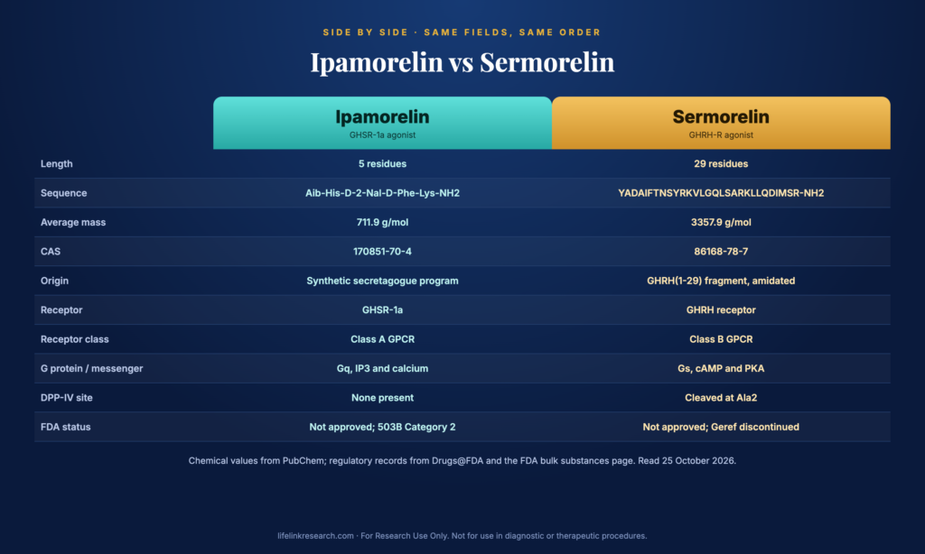
    FieldIpamorelinSermorelin
    Full nameIpamorelin (NNC 26-0161)Sermorelin, GHRH(1-29)-NH2
    SequenceAib-His-D-2-Nal-D-Phe-Lys-NH2YADAIFTNSYRKVLGQLSARKLLQDIMSR-NH2
    Length5 residues (pentapeptide)29 residues
    Molecular formulaC38H49N9O5C149H246N44O42S
    Molecular weight711.9 g/mol (PubChem CID 9831659)3357.9 g/mol (PubChem CID 16132413)
    CAS number170851-70-486168-78-7
    OriginSynthetic, from a secretagogue chemistry programN-terminal 1-29 fragment of human GHRH, amidated
    ReceptorGHSR-1a (ghrelin receptor)GHRH receptor
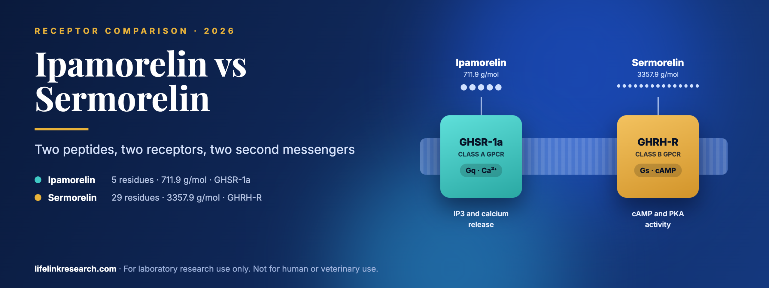
    Receptor class and signalingClass A GPCR, Gq, IP3 and calciumClass B GPCR, Gs, cAMP and PKA
    Selectivity dataAntagonist profiling in rat pituitary cells and animal models (Raun 1998)Defined by its parent hormone’s receptor
    Main assaysRadioligand binding, calcium flux, pituitary-cell readoutscAMP accumulation, pituitary-cell readouts, DPP-IV stability
    Study typesIn vitro and animalIn vitro and animal
    FDA statusNot approved; listed in Category 2 of the 503B bulks reviewNot approved as marketed today; Geref discontinued
    Life Link formatIpamorelin 10mg, lyophilized powder, $67, 44 in stockNot stocked

    The Sermorelin vs Ipamorelin difference that drives everything downstream shows up in two rows of that table. The receptor-class row decides which second-messenger assay reads out at all, and the length row decides how exposed each chain is to peptidases.

    What is Sermorelin?

    Quick answer: Sermorelin is the first 29 residues of human growth-hormone-releasing hormone with an amidated C-terminus, written GHRH(1-29)-NH2. PubChem lists C149H246N44O42S at 3357.9 g/mol, CAS 86168-78-7. It acts at the GHRH receptor, a class B GPCR that couples to Gs and raises cAMP.

    Sermorelin is the shortened version of a hormone the body already makes, which means its pharmacology was mapped before the fragment itself existed as a research tool.

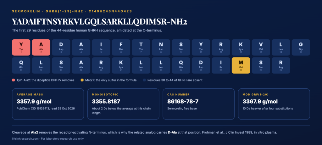
    Sermorelin GHRH(1-29) amino acid sequence

    Sermorelin GHRH(1-29) amino acid sequence

    Origin

    Human GHRH is 44 residues long. Sermorelin reproduces residues 1 to 29 and caps the chain with an amide. The reason that fragment works is positional: the receptor-activating information in GHRH is concentrated in the N-terminal region, so truncating the C-terminal 15 residues removes bulk without removing the part the receptor reads. The amide cap replaces the charged carboxyl that truncation would otherwise leave exposed.

    Chemical identity

    Sermorelin identity fieldValue
    Sequence, single-letterYADAIFTNSYRKVLGQLSARKLLQDIMSR-NH2
    Length29 residues, amidated C-terminus
    Average mass3357.9 g/mol
    Monoisotopic mass3355.8187
    Sulfur-containing residueMet27, the only S in the formula
    CAS86168-78-7

    That lone methionine at position 27 is more than a formula detail. It is an oxidation-sensitive residue, and it is also one of the four positions that structurally related analogs change, which the CJC-1295 section below returns to.

    What is Ipamorelin?

    Quick answer: Five residues, two of them non-natural: Aib-His-D-2-Nal-D-Phe-Lys-NH2 is Ipamorelin, formula C38H49N9O5, 711.9 g/mol, CAS 170851-70-4. Its target is GHSR-1a, the ghrelin receptor, which it reaches with no structural resemblance to ghrelin at all. Our Ipamorelin structure and GHSR guide covers it in full.

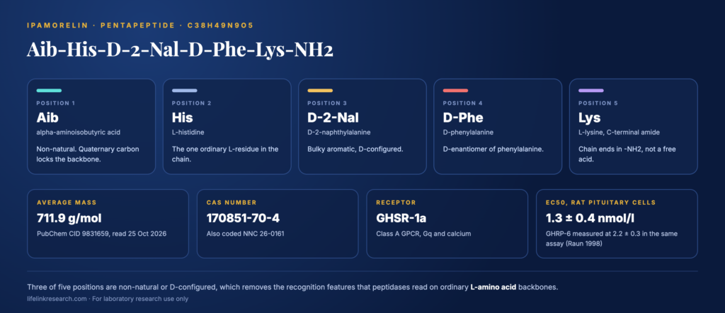
    Ipamorelin (Aib-His-D-2-Nal-D-Phe-Lys-NH2) chemical structure

    Ipamorelin (Aib-His-D-2-Nal-D-Phe-Lys-NH2) chemical structure

    Origin

    Raun and colleagues at Novo Nordisk described Ipamorelin in European Journal of Endocrinology in 1998 (PMID 9849822). It emerged from a series of compounds built by removing the central Ala-Trp dipeptide from GHRP-1, which is why it reads as a chemistry-program output and not as a natural fragment.

    Chemical identity

    Two of its five positions are non-natural, and that is deliberate design, not incidental chemistry.

    • Aib (alpha-aminoisobutyric acid), position 1: a doubly methylated alanine. Its quaternary carbon constrains the backbone and removes the usual recognition features at the N-terminus.
    • D-2-Nal, position 3: D-configured 2-naphthylalanine, a bulky aromatic residue not found in natural proteins.
    • D-Phe, position 4: the D-enantiomer of phenylalanine.
    • C-terminal amide: the chain ends in -NH2 instead of a free acid.

    GHRH receptor vs ghrelin receptor: how do the targets differ?

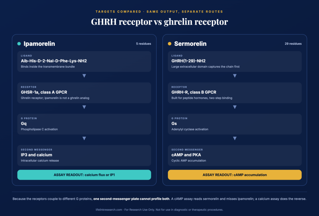
    Quick answer: The GHRH receptor is a class B GPCR that couples to Gs and raises cAMP through adenylyl cyclase. GHSR-1a is a class A GPCR that couples to Gq, activating phospholipase C to generate IP3 and release intracellular calcium. Different receptor families, different G proteins, different second messengers, and different assay plates.

    GHRH receptor and GHSR-1a signaling pathway comparison diagram

    GHRH receptor and GHSR-1a signaling pathway comparison diagram

    GHRH-R: class B GPCR, Gs, cAMP and PKA

    Class B receptors are built for peptide hormones. They carry a large extracellular domain that captures the peptide’s C-terminal portion first, then lets the N-terminal residues engage the transmembrane core. That two-step geometry explains why a 29-residue fragment still activates the receptor while a five-residue peptide never could: there has to be enough chain to occupy both contact regions.

    Downstream, the receptor couples to Gs, adenylyl cyclase produces cAMP and PKA activity follows. In practice that makes cAMP accumulation the standard readout for anything targeting GHRH-R.

    GHSR-1a: class A GPCR, Gq, IP3 and calcium

    GHSR-1a belongs to the much larger class A family, where the ligand binds within the transmembrane bundle itself, with no separate extracellular domain involved. A compact pocket of that kind can be filled by a well-shaped pentapeptide, which is why Ipamorelin’s size is not an obstacle.

    Its canonical coupling is to Gq, running through phospholipase C to IP3 and intracellular calcium release, so calcium flux or IP1 accumulation is the usual readout. Work on agonist-specific coupling at this receptor (Neuroendocrinology, 2004, PMID 14755130) found that different agonists engage its intracellular signaling systems differently, an in vitro finding worth weighing when choosing which readout to measure.

    One point deserves emphasis because secondary sources blur it: Ipamorelin is not a ghrelin analog by structure. Ghrelin is a 28-residue acylated peptide. Ipamorelin is five residues with no acyl group and no sequence relationship to it. Shared receptor, unrelated molecules.

    Selectivity data

    The 1998 paper established Ipamorelin’s receptor assignment through pharmacology rather than inference. In primary rat pituitary cells, Ipamorelin produced its readout with an EC50 of 1.3 ± 0.4 nmol/l, close to GHRP-6 at 2.2 ± 0.3 nmol/l. Antagonist profiling in that same in vitro system then showed the readout was blocked by a GHRP antagonist and not by a GHRH antagonist, which assigns the activity to the GHRP-type receptor now known as GHSR-1a.

    Selectivity was examined further in anesthetized rat and conscious swine models, where the paper compared Ipamorelin against other secretagogues across additional pituitary hormone readouts. Those are animal-model findings, reported here as study design only, with no outcomes attached.

    How stable are Ipamorelin and Sermorelin in assay conditions?

    Quick answer: Sermorelin carries the GHRH N-terminal sequence, which dipeptidyl peptidase IV cleaves after the Ala at position 2. Ipamorelin’s non-natural residues (Aib at position 1, D-amino acids at 3 and 4) remove the recognition features peptidases rely on, so it resists the same enzymatic routes.

    Frohman and colleagues (J Clin Invest, 1989, PMID 2565342) identified dipeptidylpeptidase IV and trypsin-like activity as the enzymatic routes that degrade human GHRH in plasma in vitro. The DPP-IV step removes the first two residues, Tyr-Ala, and because the N-terminal region carries the receptor-activating information, the truncated product is no longer the same ligand.

    That one cleavage site explains a great deal of the chemistry that followed:

    • Sermorelin keeps native Ala at position 2, so it keeps the DPP-IV recognition motif intact.
    • Structurally related analogs substitute D-Ala at position 2, which DPP-IV does not accept, which is the single most consequential change in the group.
    • Ipamorelin has no DPP-IV site to protect, because Aib at position 1 and D-residues at 3 and 4 are not substrates for the proteases that read natural L-amino acid backbones.

    These are in vitro enzymatic observations, not statements about duration in any organism.

    Where do CJC-1295 (no DAC) and Mod GRF(1-29) fit?

    Quick answer: Mod GRF(1-29), also sold as CJC-1295 no DAC, is GHRH(1-29) carrying four substitutions: D-Ala at position 2, Gln at 8, Ala at 15 and Leu at 27. It is structurally related to Sermorelin, not equivalent to it. The DAC version adds a linker that the no-DAC form does not have.

    Reading the registry sequence for CAS 863288-34-0 (PubChem CID 56841945) against native GHRH(1-29) confirms all four positions:

    PositionSermorelin, GHRH(1-29)Mod GRF(1-29)What the change does
    2AlaD-AlaRemoves the DPP-IV cleavage motif
    8AsnGlnReplaces a deamidation-prone residue
    15GlyAlaAdds a side chain at a flexible position
    27MetLeuRemoves the only oxidation-sensitive sulfur

    Three of those four changes target chemical liabilities rather than receptor contact, which is why the analog is described as structurally related: same receptor family, same 29-residue length, different stability profile. The mass difference follows from the substitutions, 3367.9 g/mol against Sermorelin’s 3357.9.

    Life Link stocks CJC-1295 No DAC 5mg at $38, which is the closest available material to Sermorelin for GHRH-receptor work, since Sermorelin itself is not in the catalog. For the pairing comparison, see CJC-1295 vs Ipamorelin.

    What assays and models are used to study each?

    Quick answer: Radioligand binding plus cAMP accumulation profiles for GHRH-receptor compounds. Radioligand binding plus calcium or IP1 profiles for GHSR-1a compounds. Primary pituitary cells carry both receptors, so hormone release there serves as a shared in vitro readout, and LC-MS identity confirmation precedes all of it.

    AssayIpamorelinSermorelinStudy type
    Radioligand bindingGHSR-1a membranesGHRH-R membranesIn vitro
    cAMP accumulationNot the primary readoutPrimary readout (Gs-coupled)In vitro
    Calcium flux or IP1Primary readout (Gq-coupled)Not the primary readoutIn vitro
    GH release from primary pituitary cellsReported, EC50 1.3 nmol/l (Raun 1998)Reported for GHRH-R agonistsIn vitro readout
    DPP-IV stabilityNo cleavage motif presentCleaved at Ala2In vitro, plasma
    LC-MS identityExpected mass near 711.9Expected mass near 3357.9Analytical

    Tracer choice separates the two workflows as well. A GHSR-1a binding experiment runs against a labeled secretagogue ligand, while a GHRH-receptor experiment runs against a labeled GHRH-type peptide, so the two assays share no reference compound, and their numbers are not comparable across plates. Identity confirmation comes first in both cases, because a binding curve generated from mislabeled material is simply a curve for whatever was actually in the vial.

    Two design points follow. Because the receptors couple to different G proteins, a single second-messenger plate cannot profile both compounds: a cAMP assay will read Sermorelin and miss Ipamorelin, and a calcium assay does the reverse. And because primary pituitary cells express both receptors, that system is the one place the two converge, which makes antagonist controls the only way to attribute a readout to one receptor or the other, exactly as the 1998 work did.

    Can Ipamorelin and Sermorelin be studied together?

    Quick answer: Co-incubation is feasible in primary pituitary cells, which carry both receptors, and the published secretagogue literature has examined GHRH-type and GHRP-type compounds in the same preparation. The design requirement is antagonist controls, since two receptors converge on one measurable output.

    Precedent for the design already exists in the founding literature. The 1998 antagonist profiling was itself a co-incubation experiment: Ipamorelin was applied to primary rat pituitary cells alongside a GHRH antagonist in one condition, and a GHRP antagonist in another, and the contrast between those two conditions is what assigned the receptor. Published work pairing a GHRH-type compound with a GHRP-type compound in the same preparation follows the same logic.

    Beyond the combination well itself, a dual-receptor design calls for single-compound conditions matched on plate and passage, and a receptor-selective antagonist arm covering each pathway. Skip the antagonist arms and a combined readout can be attributed to neither receptor.

    Two further points come from the chemistry above. The compounds differ 4.7-fold in mass, so equal-mass preparation and equal-molar preparation describe different experiments, and the protocol should state which. And since the two readouts run through different second messengers, a combination study usually needs two measurement systems in parallel. Life Link supplies the composition pairing as CJC-1295 + Ipamorelin 5/5mg at $72, which pairs the Ipamorelin pentapeptide with the GHRH-type analog, since Sermorelin itself is not stocked.

    What is the FDA status of Ipamorelin and Sermorelin?

    Quick answer: Neither compound is an FDA-approved product today. Sermorelin acetate was approved as Geref under NDA 019863 on December 28, 1990, and as Geref Diagnostic under NDA 020443 on September 26, 1997; both are recorded as Discontinued. Ipamorelin acetate appears in Category 2 of the FDA’s 503B bulk substances review.

    The Sermorelin approvals and their discontinued marketing status come from the FDA’s own Drugs@FDA records, with EMD Serono as the listed sponsor. That is a record of marketing history only.

    For Ipamorelin, the FDA’s page on bulk drug substances that may present significant safety risks lists Ipamorelin acetate in Category 2, added September 29, 2023, under the 503B outsourcing-facility review. FDA stamps that page as current to April 22, 2026.

    Three points keep that status accurate:

    • Category 2 here concerns compounded preparations made by outsourcing facilities. It is a separate question from the supply of research reagents to laboratories.
    • A discontinued approval is not a withdrawal for cause, and this page makes no statement about why marketing ended.
    • Neither compound is approved for any use as of October 25, 2026.

    Identity and quality checks for research supply

    A 4.7-fold mass difference makes these two easy to separate analytically. The documentation still has to be read in a particular order.

    • Purity: purity by HPLC, so see the batch result for the figure that applies to the vial you hold, and check the batch number on the report against the label.
    • LC-MS identity: expected values are near 711.9 for Ipamorelin and near 3357.9 for Sermorelin. Mod GRF(1-29) lands at 3367.9, only 10 daltons from Sermorelin, which is the one pair on this page where an identity trace has to be read carefully. Walking a certificate line by line is covered in how to read a peptide COA.
    • Form and storage: lyophilized powder, listed at -20°C with protection from light.
    • Solubility: described as soluble in water or DMSO for assay preparation.

    Mass convention needs stating at this size range too. For the 711.9 g/mol pentapeptide, the average and monoisotopic figures sit about 0.5 Da apart, so either one identifies the compound unambiguously. For a 3.3 kDa chain like Sermorelin the separation widens to roughly 2 Da, and the isotope envelope no longer peaks at the monoisotopic mass, so a report quoting one convention against a reference using the other can look like a failure when the material is correct.

    For Sermorelin specifically, the Met27 residue makes oxidation a sensible thing to watch on an identity trace, since a +16 increase on a single methionine is the classic signature.

    FAQs

    What is the difference between Ipamorelin and Sermorelin?

    Ipamorelin is a five-residue synthetic peptide that binds GHSR-1a, a class A GPCR signaling through Gq and calcium. Sermorelin, the 29-residue GHRH(1-29)-NH2 fragment, binds a class B GPCR, the GHRH receptor, which signals through Gs and cAMP. Different receptors, different second messengers.

    Do Ipamorelin and Sermorelin act on the same receptor?

    No. Ipamorelin acts at GHSR-1a, the ghrelin receptor, and Sermorelin at the GHRH receptor. Raun and colleagues established Ipamorelin’s assignment in 1998 using antagonist profiling in rat pituitary cells: a GHRP antagonist blocked the readout, while a GHRH antagonist did not.

    Is Sermorelin a GHRH analog?

    Yes. Sermorelin is GHRH(1-29)-NH2, meaning it reproduces the first 29 residues of the 44-residue human GHRH sequence with an amidated C-terminus. Those residues carry the receptor-activating region, which is why the shortened chain still binds the GHRH receptor.

    What is the difference between GH and GHRH?

    They are different molecules at different points of the same axis. Growth hormone is a 191-residue protein. GHRH is a 44-residue peptide that binds its own receptor on pituitary cells. Sermorelin corresponds to GHRH’s first 29 residues, not to growth hormone.

    Is CJC-1295 the same as Sermorelin?

    No. CJC-1295 no DAC, also called Mod GRF(1-29), is GHRH(1-29) with four substitutions: D-Ala2, Gln8, Ala15 and Leu27, confirmed against PubChem’s registry sequence. The DAC version also carries a linker. Sermorelin has none of these changes.

    What receptor does Ipamorelin bind?

    Its target is GHSR-1a, known both as the ghrelin receptor and as growth hormone secretagogue receptor type 1a. In primary rat pituitary cells its EC50 was 1.3 ± 0.4 nmol/l, comparable to GHRP-6, with selectivity confirmed by antagonist profiling in that in vitro system and examined further in animal models.

    Is Sermorelin FDA approved?

    Not as a marketed product today. Sermorelin acetate was approved as Geref (NDA 019863) on December 28, 1990, and as Geref Diagnostic (NDA 020443) on September 26, 1997. Drugs@FDA records both as Discontinued, checked October 25, 2026.

    Ipamorelin vs Sermorelin comparison table: receptor, class, signaling, sequence and mass

    Ipamorelin vs Sermorelin comparison table: receptor, class, signaling, sequence and mass

    Closing notes

    For Research Use Only. Not for use in diagnostic or therapeutic procedures. Read our full Research Use Disclaimer.

    Materials described here are laboratory reagents supplied to qualified researchers. The full research-use policy is set out in the Terms and Conditions.

    Author: Trevor Blackwell. Scientific reviewer: Danielle Reyes, PhD (receptor pharmacology). This page was last reviewed on October 25, 2026, the date every chemical value here was read from PubChem and every regulatory record from FDA sources.

    References

  • KPV vs BPC-157: Molecular Identity, Pathways and Research Compared

    KPV vs BPC-157: Molecular Identity, Pathways and Research Compared

    A PubMed search run in late October returns 230 records for BPC-157 and 47 for KPV. Exactly one record names both, and it is a 2026 pharmacy-compounding policy review, not a laboratory study. Two molecules that keep appearing in the same catalogs, the same forum threads, and the same regulatory agendas have almost no shared literature behind them. They also have almost nothing in common chemically: one is three residues long, the other fifteen, and roughly 1,077 daltons separate them. This guide compares KPV vs BPC-157 on identity and research tooling only.

    Sequence, formula, mass, CAS number, origin, the receptors and transporters each has been studied against, the assay types that generate the published readouts, and the documentation a receiving lab uses to confirm which vial holds which compound.

    Quick Answer

    KPV is a 3-amino-acid fragment (Lys-Pro-Val) of alpha-MSH, studied with PepT1 uptake and NF-κB signaling assays. BPC-157 is a synthetic 15-amino-acid peptide studied in VEGFR2, eNOS, and FAK-paxillin pathway assays. Both have mostly in vitro and rodent data, and neither is FDA-approved.

    KPV vs BPC-157 at a glance

    Quick answer: KPV is a tripeptide of 342.43 g/mol cut from the C-terminus of alpha-MSH. BPC-157 is a synthetic pentadecapeptide of 1419.5 g/mol whose sequence comes from a protein isolated from gastric juice. KPV is studied through a peptide transporter; BPC-157 through receptor-expression and kinase readouts with no single identified receptor.

    Every chemical value below was read from PubChem on October 25, 2026. Format and stock figures come from the Life Link Research catalog on the same date.

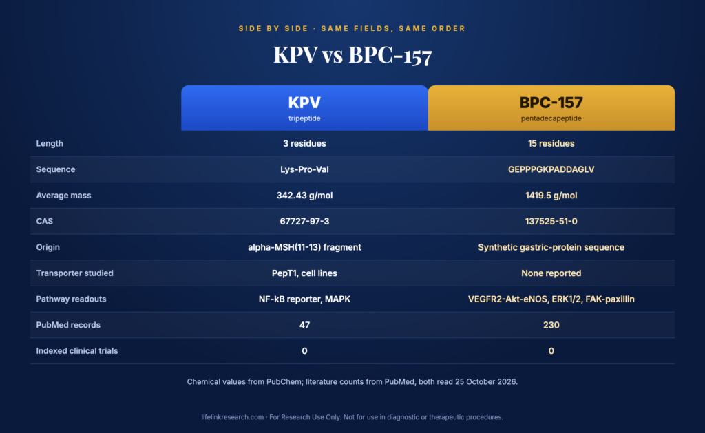
    FieldKPVBPC-157
    Full nameLys-Pro-Val, alpha-MSH(11-13)Body Protection Compound 157 (also coded PL 14736)
    SequenceLys-Pro-ValGly-Glu-Pro-Pro-Pro-Gly-Lys-Pro-Ala-Asp-Asp-Ala-Gly-Leu-Val (GEPPPGKPADDAGLV)
    Length3 residues (tripeptide)15 residues (pentadecapeptide)
    Molecular formulaC16H30N4O4C62H98N16O22
    Molecular weight342.43 g/mol average, 342.2267 monoisotopic (PubChem CID 125672)1419.5 g/mol average, 1418.7042 monoisotopic (PubChem CID 9941957)
    CAS number67727-97-3137525-51-0
    Where it comes fromC-terminal fragment of the hormone alpha-MSH, residues 11 to 13Synthetic partial sequence of a protein first isolated from gastric juice
    Receptors and transporters studiedPepT1 (di/tripeptide transporter), measured in cell linesNo single identified receptor; VEGFR2 and growth-hormone-receptor expression reported as readouts
    Main assay typesNF-κB reporter, uptake with competitor, cytokine ELISATube formation, migration, Western blot for kinase phosphorylation, qPCR
    Main study typesIn vitro and rodentIn vitro and rodent
    Human data statusNo PubMed records indexed as clinical trials (checked October 25, 2026)No PubMed records indexed as clinical trials (checked October 25, 2026)
    Life Link formatKPV 10mg, lyophilized powder, $97, 21 in stockBPC-157 10mg, lyophilized powder, $89, 12 in stock

    Two numbers in that table carry most of the practical difference. The 4.1-fold mass ratio decides what a mass spectrometer should show, and the 3-versus-15 residue count decides how much chain there is for enzymes to cut.

    What is KPV?

    Quick answer: KPV is the tripeptide Lys-Pro-Val, occupying the final three positions of alpha-melanocyte-stimulating hormone. PubChem lists it as C16H30N4O4 with an average mass of 342.43 g/mol and CAS 67727-97-3. In published cell work it is taken up through the PepT1 transporter and assayed with NF-κB reporter constructs.

    Short peptides give a lab a defined, inexpensive molecule with very few variables. KPV takes that to an extreme: three residues, one basic side chain, one proline and one branched aliphatic residue. There is no secondary structure to speak of and no disulfide chemistry to manage.

    KPV (Lys-Pro-Val) chemical structure

    KPV (Lys-Pro-Val) chemical structure

    Where it comes from

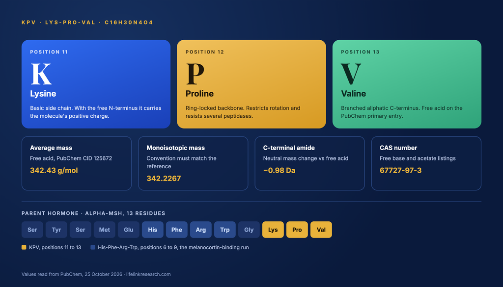
    Alpha-MSH is a 13-residue hormone. KPV is its last three residues, written in the literature as alpha-MSH(11-13). The portion of alpha-MSH that binds melanocortin receptors is the His-Phe-Arg-Trp run at positions 6 to 9, which falls outside the KPV fragment entirely. That single structural point explains why KPV behaves in assays the way a transporter substrate does, and not the way a receptor ligand does, and it is the detail most comparison pages leave out. Our KPV research guide covers the parent hormone in more depth.

    Chemical identity

    PubChem’s primary KPV entry, CID 125672, describes the free acid, meaning the C-terminal valine ends in a carboxyl group. That distinction is not cosmetic. Swapping the terminal hydroxyl for an amide lowers the neutral mass by about 0.98 Da, so a C-terminally amidated preparation and a free-acid preparation produce different expected ions on the same instrument. Anyone matching an observed mass against a reference value needs to know which form the reference describes, and whether the figure quoted is average or monoisotopic. At this molecular size those two conventions differ by roughly 0.2 Da.

    KPV identity fieldValue
    C-terminus on the PubChem primary entryFree acid
    Average mass, free acid342.43 g/mol
    Monoisotopic mass, free acid342.2267
    Expected [M+H]+ , average343.4
    Net charge at neutral pH, free acidApproximately +1

    What is BPC-157?

    Quick answer: BPC-157 is a synthetic 15-residue peptide with the sequence GEPPPGKPADDAGLV. PubChem puts the formula at C62H98N16O22, the average mass at 1419.5 g/mol and the CAS number at 137525-51-0. Published work uses endothelial and fibroblast cell systems with VEGFR2, eNOS and FAK-paxillin readouts, plus rodent models.

    Where KPV is a hormone fragment, BPC-157 is a laboratory construct. It is assembled by solid-phase synthesis and has no single natural precursor sold alongside it, which makes the sequence itself the whole of its identity.

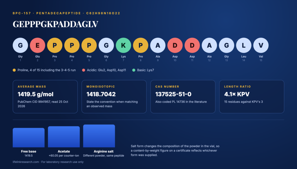
    BPC-157 (GEPPPGKPADDAGLV) 15-residue sequence diagram

    BPC-157 (GEPPPGKPADDAGLV) 15-residue sequence diagram

    Where it comes from

    The sequence was described as a partial sequence of a protein first isolated from gastric juice. That statement describes the origin of the amino acid order and nothing else: the molecule supplied to labs today is made synthetically, not extracted. Three of its fifteen positions are prolines in a row (positions 3, 4 and 5), with a fourth at position 8, and that proline content is the structural feature most often cited when researchers discuss its behavior in aqueous buffers. The BPC-157 research guide goes through the sequence position by position.

    Salt forms

    BPC-157 appears in catalogs as a free base, an acetate salt and an arginine salt. These are identity facts, not performance claims, and each changes the weight of the powder in the vial without changing the peptide.

    • Free base: the peptide alone, 1419.5 g/mol.
    • Acetate: each acetate counter-ion adds about 60.05 g/mol. BPC-157 has basic sites at Lys7 and the N-terminus, so a fully acetated preparation runs roughly 8% heavier than the free base.
    • Arginine salt: arginine serves as the counter-ion instead. Catalogs that list an arginine-salt product are describing a different powder composition, not a different peptide.

    A certificate reporting peptide content by weight will reflect whichever form is in the vial, which is why the salt form belongs on the label.

    How do KPV and BPC-157 differ at the molecular level?

    Quick answer: KPV is small enough to be a substrate for a peptide transporter and has a documented route into cells. BPC-157 is four times heavier, has no single identified receptor in the published literature, and is studied through downstream kinase and receptor-expression readouts in place of direct binding.

    Receptors and transporters

    KPV has the clearer entry route of the two. Working in Caco2-BBE and HT29-Cl.19A cell lines, Dalmasso and colleagues (Gastroenterology, 2008, PMID 18061177) reported that “KPV is transported into cells by PepT1,” the di- and tripeptide transporter, and demonstrated it with uptake experiments using unlabeled KPV as a competitor. That is an in vitro finding in human cell lines.

    KPV vs BPC-157 comparison table graphic: sequence, mass, origin, pathways and study types

    KPV vs BPC-157 comparison table graphic: sequence, mass, origin, pathways and study types

    Whether KPV engages melanocortin receptors is a point where secondary sources conflict and the primary literature does not support the stronger claim. No peer-reviewed binding measurement for KPV at MC1R or MC3R appeared in our October 2026 search. Land (Int J Physiol Pathophysiol Pharmacol, 2012, PMID 22837805), frequently cited as evidence for MC3R involvement, attributed the MC3R requirement to gamma-MSH and traced KPV’s measured activity in 16HBE14o- bronchial epithelial cells to its own nuclear import and competition at the importin-alpha binding site on p65RelA. Combined with the absence of the His-Phe-Arg-Trp pharmacophore, the published record points away from receptor binding as KPV’s assay mechanism.

    BPC-157 has no equivalent entry point on record. No receptor has been identified as its binding partner in the published literature, so studies report expression and activation of other proteins as readouts. Hsieh and colleagues (J Mol Med, 2017, PMID 27847966) measured VEGFR2 expression and internalization in endothelial cells, with the dynamin inhibitor dynasore used as a block. Separately, Chang and colleagues (Molecules, 2014, PMID 25415472) reported growth-hormone-receptor expression in rat tendon fibroblasts by Western blot and RT-PCR. Both are readouts of what a cell does in the compound’s presence, not demonstrations of a binding site.

    Signaling pathways measured

    Pathway or readoutKPVBPC-157
    NF-κB reporter activityPrimary readout, luciferase constructsNot a standard readout
    MAPK phosphorylationReported alongside NF-κBReported as ERK1/2
    VEGFR2 to Akt to eNOSNot studiedPrimary readout
    FAK and paxillin phosphorylationNot studiedReported in fibroblast work
    Transporter-mediated uptakePepT1, with competitorNot reported

    The two columns barely overlap, which is the most useful thing a comparison can tell a lab planning controls.

    What assays and models are used to study each?

    Quick answer: KPV work centers on transporter uptake and transcription-factor reporter assays in epithelial and immune cell lines. BPC-157 work centers on endothelial and fibroblast assays measuring migration, tube formation and kinase phosphorylation. Both literatures are dominated by in vitro experiments and rodent models, with no indexed clinical trials for either.

    AssayKPVBPC-157Study type
    PepT1 uptake with competitorReported, Caco2-BBENot reportedIn vitro
    NF-κB luciferase reporterPrimary readoutNot standardIn vitro
    Cytokine ELISA readoutsReported in cell supernatantsOccasionally reportedIn vitro
    Cell migration and scratch assaysRarely usedCommon, fibroblasts and endothelial cellsIn vitro
    Tube formationNot reportedCommon, endothelial cellsIn vitro
    qPCR receptor expressionRarely usedCommon (VEGFR2, GH receptor)In vitro and rodent tissue
    Stability in culture mediaShort chain, few cleavage sitesReported as stable in aqueous bufferIn vitro

    Three practical consequences follow from that table. KPV’s assay set depends on a transporter being present and functional, so a cell line’s PepT1 expression is a design variable, so it belongs in the methods section. BPC-157’s assay set is mostly phenotypic, which puts more weight on vehicle controls and on blinding during image analysis. And because neither compound has a reference antagonist in common use, specificity in both cases rests on the control conditions a lab builds, since no pharmacological block is available.

    How much published research exists on KPV vs BPC-157?

    Quick answer: As of October 25, 2026, PubMed indexes 230 records for BPC-157 and 47 for KPV. Neither compound has a record indexed as a clinical trial. 108 BPC-157 records and 13 KPV records carry the animals indexing tag without the humans tag, and reviews account for 51 and 8 records respectively.

    Measure, PubMed, October 25, 2026KPVBPC-157
    Total records, title and abstract47230
    Indexed as review851
    Animal-indexed, not human-indexed13108
    Indexed as clinical trial or RCT00
    Records naming both compounds1 (a 2026 policy review)1 (the same record)

    Anyone weighing BPC-157 vs KPV for research planning should read those counts carefully, because two features of them shape what the totals actually mean. BPC-157’s total is nearly five times KPV’s, but more than a fifth of it is review articles, so the primary-study difference is narrower than the headline figures suggest.

    And the zero in the clinical-trial row is the same zero for both: human data are limited, and nothing in either literature is indexed as a controlled trial. The KPV query also required a peptide or melanocortin term, because the bare initials collide with unrelated abbreviations across the database.

    For a comparison built on a similar count-first approach, see BPC-157 vs TB-500.

    Can KPV and BPC-157 be studied together?

    Quick answer: Combined in vitro work is possible but has essentially no published precedent. One PubMed record names both compounds as of October 25, 2026, and it is a regulatory review with no experiments in it. Any combined design is therefore building its own baseline from scratch.

    That single overlapping record (Willis and Vu, Int J Pharm Compd, 2026, PMID 42752426) analyzes compounding-committee recommendations. It reports no assay data for either molecule.

    The absence of precedent raises the bar for controls. A combined-exposure plate needs four arms at minimum: vehicle, each compound alone at its own concentration series, and the combination. Without the single-compound arms run on the same plate and the same passage, nothing in the combination well can be assigned to either molecule.

    Two further design points follow from the chemistry already described. Because KPV’s uptake depends on PepT1, any combination assay should verify that transporter expression is unchanged across conditions. And since the two compounds differ more than fourfold in mass, an equal-mass exposure delivers roughly four times as many KPV molecules as BPC-157 molecules, so molar matching and mass matching give different experiments. State which one the protocol uses.

    What is the FDA status of KPV and BPC-157?

    Quick answer: As of October 25, 2026, neither KPV nor BPC-157 is an FDA-approved drug. Neither appears in the FDA’s approved-products database. Both were reviewed by the Pharmacy Compounding Advisory Committee on July 23, 2026, in free base and acetate forms, as candidates for the 503A bulk drug substances list.

    The FDA’s advisory committee calendar entry for the July 23 to 24, 2026 meeting lists “BPC-157 (free base)”, “BPC-157 acetate”, “KPV (free base)” and “KPV acetate” among the substances considered on the first day. The page was marked content current as of August 6, 2026.

    Three points of context keep that status accurate:

    • An advisory committee recommendation is advisory. Placement on the 503A list requires FDA action through notice-and-comment rulemaking, and a committee vote is not an approval.
    • The 503A list governs what compounding pharmacies may use. It is a separate question from the sale of research reagents to laboratories.
    • Neither compound appears as an approved product in the FDA’s drug approval records, searched on October 25, 2026.

    Our BPC-157 regulatory status page tracks the list itself, and the 2026 FDA advisory panel write-up covers the July meeting in detail.

    Identity and quality checks for research supply

    Because the two molecules differ so widely in mass, the documentation that distinguishes them is straightforward to read once you know which three fields to compare.

    • Purity by HPLC: reverse-phase HPLC with UV detection expresses how much of the sample is the named peptide, as an area percentage. See the batch result, which supersedes any catalog figure, and check that the report’s batch number matches the vial.
    • Mass by LC-MS: the measured mass needs to line up with the figure predicted for that compound in that form. For KPV, that means a value near 342.4 for the free-acid free base, and for BPC-157 near 1419.5. A salt form changes the powder’s composition, so confirm which the report describes before calling a figure a mismatch. For the certificate layout itself, how to read a peptide COA sets out which box holds which number.
    • Form and storage: both compounds are supplied as lyophilized powder and listed for storage at -20°C, protected from light.
    • Solubility: both are described as soluble in water or DMSO for assay preparation.

    A final identity note specific to this pair. A 342-dalton peak and a 1,419-dalton peak cannot be confused with one another, but a KPV report quoting a monoisotopic value against an average-mass reference can look wrong when it is not. Match convention to convention.

    FAQs

    What is the difference between KPV and BPC-157?

    KPV is three residues long (Lys-Pro-Val) and comes from positions 11 to 13 of alpha-MSH. BPC-157 is fifteen residues (GEPPPGKPADDAGLV) and is a synthetic sequence from a gastric-juice protein. KPV is studied via PepT1 uptake and NF-κB readouts; BPC-157 via VEGFR2, eNOS and FAK-paxillin readouts.

    What is KPV’s full name?

    KPV stands for its three amino acids: lysine, proline and valine, written Lys-Pro-Val. In the literature it is also written alpha-MSH(11-13), because the three residues correspond to positions 11 to 13 of alpha-melanocyte-stimulating hormone. Its CAS number is 67727-97-3.

    What is the molecular weight of KPV?

    PubChem lists KPV at an average molecular weight of 342.43 g/mol, with a monoisotopic mass of 342.2267, for the formula C16H30N4O4 (CID 125672, read October 25, 2026). That entry describes the free acid. An amidated C-terminus lowers the neutral mass by roughly 0.98 Da.

    What is the BPC-157 peptide sequence?

    BPC-157 is Gly-Glu-Pro-Pro-Pro-Gly-Lys-Pro-Ala-Asp-Asp-Ala-Gly-Leu-Val, written in single-letter code as GEPPPGKPADDAGLV. That is fifteen residues, which is why it is described as a pentadecapeptide. PubChem lists the formula as C62H98N16O22 at 1419.5 g/mol average mass.

    Is there a lot of research on BPC-157?

    PubMed indexed 230 BPC-157 records on October 25, 2026, of which 51 are reviews, and 108 carry the animals tag without a humans tag. No record is indexed as a clinical trial or randomized controlled trial, so the literature is predominantly in vitro and rodent work, and human data are limited.

    Can KPV and BPC-157 be studied together?

    Only one PubMed record names both, and it is a 2026 compounding-policy review with no assay data. A combined in vitro design therefore needs each compound alone on the same plate as controls, plus a stated choice between molar matching and mass matching, since the two differ fourfold in mass.

    Is KPV or BPC-157 FDA approved?

    Neither is FDA-approved. As of October 25, 2026, neither appears in the FDA’s approved drug records. Both were reviewed by the Pharmacy Compounding Advisory Committee on July 23, 2026, in free base and acetate forms, as candidates for the 503A bulk substances list. A committee review is not an approval.

    Closing notes

    For Research Use Only. Not for use in diagnostic or therapeutic procedures. Read our full Research Use Disclaimer.

    The products described on this page are laboratory reagents sold to qualified researchers. See the Terms and Conditions for the full policy. Chemical values read from PubChem and literature counts pulled from PubMed on that date.

    References