• Order within –h –m
  • Free shipping on orders over $250
  • Free Bacteriostatic Water 30ml with $500+ purchase
  • Every batch tested by an independent lab
  • For laboratory and research use only. Not for human consumption

Category: Research Guides

  • How to Verify a Peptide COA Is Real (and Spot a Faked One)

    How to Verify a Peptide COA Is Real (and Spot a Faked One)

    Quick Answer

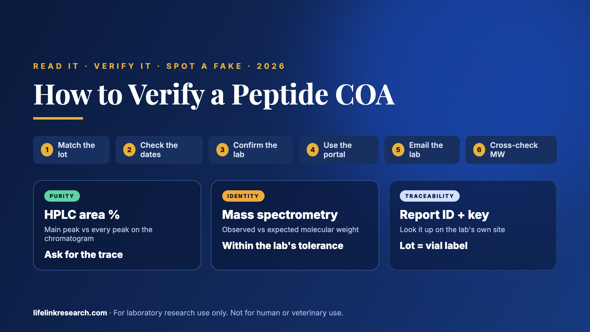
    To verify a peptide COA, check the vial’s lot number against the report, confirm the named lab exists and is accredited, look the report up on the lab’s own portal, and make sure the HPLC purity and mass-spec results fit the compound. A report that passes all four checks traces back to a real test of that batch.

    Key takeaways

    • A COA covers one batch: the lot number on the report must match the vial label
    • HPLC gives purity; mass spectrometry confirms identity by observed vs expected molecular weight
    • A real lab is findable, accredited (ISO/IEC 17025) and confirms its own report IDs
    • Most fakes are real reports with an edited name, lot or date, or one report reused across batches
    • A missing panel, such as endotoxin, means that result is unknown

    Two peptide certificates can carry the same green logo, the same layout and the same 99% figure, and only one of them traces back to a real test. That report, the peptide certificate of analysis (COA), covers a single batch, and it’s the only document that ties the vial on a research bench to an identity and purity result.

    This guide covers each step, plus:

    • What a peptide COA contains
    • How to read one line by line
    • Six checks that confirm it’s real
    • The signs of an edited or reused report
    • What no COA can show
    • A printable 10-point checklist

    What Is a Peptide Certificate of Analysis?

    Quick answer: A peptide certificate of analysis (COA) is a lab report for one specific batch. It shows the compound’s identity (usually by mass spectrometry), its purity (usually by HPLC), the lot number, the test date, and the lab that ran the test. A COA describes the tested sample only, which is one batch of many.

    Think of it as the lab’s receipt. It records who tested what, when they tested it, and what they found. Each field answers a different question:

    COA fieldWhat it tells you
    Lab name and report IDWho ran the test, plus the unique number used to look the report up
    Compound and lotThe peptide tested and the batch it came from. The lot links the report to a vial label
    Test dateWhen the analysis ran, so it can be lined up with the batch
    HPLC purity %The share of the sample’s detected signal that comes from the target peptide
    MS observed vs expected MWWhether the measured molecular weight fits the compound’s known weight (identity)
    Net peptide contentHow much of the sample’s weight is peptide once water and salts are counted
    Optional panelsExtra tests such as endotoxin, heavy metals or residual solvents

    A report with blank fields is answering fewer of those questions, and the next section shows where each answer appears on the page.

    How to Read a Peptide COA, Line by Line

    Quick answer: Read a peptide COA from top to bottom. Start with the lab name, report ID, and test date. Then check the compound name and lot number, the HPLC purity percentage with its chromatogram, and the mass-spec result comparing observed to expected molecular weight. Finish with any extra panels such as net peptide content or endotoxin.

    Header: Lab, Report ID, Dates

    The top block tells you who did the work: the lab’s name, its contact details, and a report or task number. Most reports also carry two or three dates, usually when testing was ordered, when the sample arrived, and when the analysis ran. On our current BPC-157 report from Janoshik, the sample arrived on 17 April 2026, and the analysis ran three days later, on 20 April. All of them should come before the batch was listed for sale.

    Sample Details: Compound, Lot, Quantity

    Next comes the sample itself. Look for the compound name, the client, the batch or lot number, and the labelled amount, such as 10 mg. Write the lot number down, because every later check leans on it. If a label reads BPC-157 10 mg, batch 031826, the report should say the same thing, character for character.

    HPLC Purity and the Chromatogram

    HPLC (high-performance liquid chromatography) pushes the sample through a column and records each component as a peak on a chart called a chromatogram. Purity is reported as area %: the main peak’s area divided by the total area of every peak. A clean result shows one tall main peak at a steady retention time (the minute it leaves the column), with only small peaks beside it. A percentage printed with no chromatogram, method, or wavelength gives you much less to check, so ask for the trace. For the difference a single point makes, see what 98% vs 99% purity means.

    Mass Spec: Observed vs Expected Molecular Weight

    Purity tells you how much of the sample is one thing. Mass spectrometry (MS) tells you what that thing is. The instrument measures the molecule’s mass, and the report sets the observed value beside the expected molecular weight from the compound’s formula. The two should agree within the tolerance the lab states, typically within about one dalton for a peptide of this size. HPLC alone can’t confirm identity, since a different peptide could also give one clean peak. A genuine report carries both numbers, because HPLC and mass spectrometry cannot substitute for one another.

    Net Peptide Content, Water and Counter-Ions

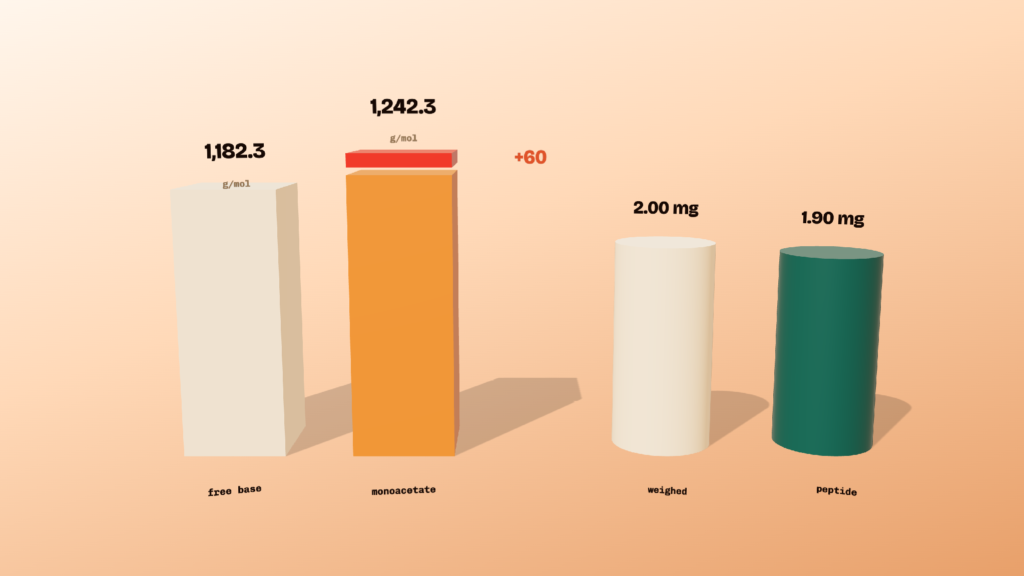
    A freeze-dried peptide sample holds more than peptide. It also carries some water and counter-ions such as acetate or TFA (trifluoroacetate) left over from synthesis and purification. Net peptide content is the share of total weight that is the peptide itself. So a sample can read 99% pure by HPLC and still contain less peptide by weight than the total weighed out. Some labs report a measured amount instead, like the 11.96 mg on our BPC-157 report for a 10 mg vial.

    Optional Panels: Endotoxin, Heavy Metals, Residual Solvents

    Extra panels are separate tests run on the same batch. Endotoxin testing uses the LAL method and reports bacterial endotoxin in EU (endotoxin units). Heavy metals testing, usually ICP-MS, measures elements such as lead, arsenic, cadmium, and mercury. Residual solvents testing, usually gas chromatography, measures solvent left from manufacturing. Each panel should show a measured value and the limit it was checked against, so read both.
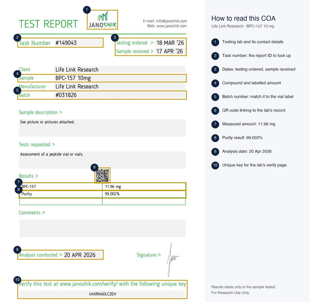
    A current Life Link Research batch COA (BPC-157 10 mg, Janoshik task #149043) with each field numbered.

    A current Life Link Research batch COA (BPC-157 10 mg, Janoshik task #149043) with each field numbered.

    How to Verify a Peptide COA Is Real: 6 Checks

    Quick answer: To verify a peptide COA, check six things. The lot number matches the vial label. The test date fits the batch. The lab exists and is accredited. The report verifies on the lab’s own portal. The lab confirms the report ID when contacted. The molecular weight matches the compound’s published formula.

    1. Match the Lot Number to the Vial Label

    Put the vial next to the report and confirm the batch number on the label matches the lot number on the COA exactly. A COA for a different lot describes a different production run, however good its numbers look. Watch for the small mismatches as well: a missing digit, a swapped prefix, or a lot that appears on the website but nowhere on the label. A vial with no lot number gives the report nothing to attach to.

    2. Check the Test Date Against the Batch

    Confirm the test date falls after the batch was made and before it went on sale. A report dated well before the batch existed can’t describe it. Watch the order of the dates inside the report too. An analysis date earlier than the sample-received date, or a date in the future, calls for a question. Stock turns over, so a years-old report attached to a newly listed product deserves one as well.

    3. Confirm the Lab Exists and Is Accredited (ISO/IEC 17025)

    Look the lab up independently and confirm it has a working website, a physical address and contact details that match the report. Then check its accreditation. ISO/IEC 17025 is the international standard for testing laboratories, and ISO says it “enables laboratories to demonstrate that they operate competently and generate valid results” (ISO).

    Accreditation comes from a recognized body, such as A2LA or ANAB in the US, and each one keeps a searchable directory of the labs it accredits. The ILAC signatory search lists recognized bodies country by country. Check the scope as well, since accreditation covers specific methods. A lab without accreditation can still produce sound data; you simply have one less outside check.

    4. Use the Lab’s Verification Portal, Key or QR Code

    Enter the report’s verification key, report ID or QR code on the lab’s own website and compare the record with your PDF. Many independent labs offer this, and it’s the strongest check you can run without picking up the phone. Here’s the process:

    1. Find the key, QR code or report ID on the COA, often near the bottom of the page.
    2. Type the lab’s web address yourself and skip any link printed inside the PDF.
    3. Enter the key or scan the QR code to open the lab’s own record.
    4. Compare that record with your copy: compound, lot, dates, and every result.

    If the portal shows a different compound, a different number, or no record at all, the PDF you have differs from what the lab issued.

    5. Email the Lab With the Report ID

    When there’s no portal, or the portal result is unclear, ask the lab directly. Send the report ID, the compound name, and the lot number, attach the PDF, and ask one question: did you issue this report with these results? Use the contact details on the lab’s own website. A written yes from the lab is about as solid as verification gets.

    6. Cross-Check the Numbers

    Test the report against public data and against itself. Start with identity: look up the compound’s formula and molecular weight in a public database such as PubChem. BPC-157, for example, is listed as C62H98N16O22 with a molecular weight of 1419.5 g/mol (PubChem CID 9941957). The expected mass on the COA should match that reference, and the observed mass should land close to it. Then compare the chromatogram with the stated purity. A 99% result should show one dominant peak with small neighbours, while a trace crowded with large peaks fits a lower number. Finally, check that the peak areas in any results table add up to roughly 100%.

    How Are Peptide COAs Faked?

    Quick answer: Fake peptide COAs are usually real reports that have been edited or reused. Common signs: a changed compound name or lot number in a PDF, one chromatogram reused across products, a single COA covering many batches, copied lab logos, dates that don’t fit the batch, and PDF metadata that shows recent editing.

    That’s useful to know, because an altered report still points back to an original record at the lab, and the edit rarely survives a side-by-side comparison. These are the signs to look for:

    Red flagHow to check it
    Edited PDF text or fontsZoom in on the compound name, lot and results. Mixed fonts, uneven spacing or a blurred patch around one field point to editing. Open the PDF’s document properties and look for a recent modified date or editing software
    Reused chromatogramCompare traces across a supplier’s products. Identical peak shapes, retention times and baseline noise on reports for different compounds can’t all be right
    One COA for many lotsCheck whether one report ID appears across several batches or products. Each batch needs its own report
    Logo with no verifiable labSearch the lab name independently. With no website, address or accreditation listing, nobody can confirm the report
    Impossible datesLook for analysis dates before sample receipt, dates in the future, or tests run before the batch existed
    MW that doesn’t match the compoundCompare the expected mass with a public database entry for the compound’s formula
    No report IDWithout an ID or key, the lab has nothing to look up. Ask the supplier for one

    A single red flag is a reason to ask questions. Two or more, and the report is best treated as unverified until the lab confirms it.

    What Can’t a COA Tell You?

    Quick answer: A COA describes only the sample the lab tested on that date. It can’t prove every vial in a batch is identical, that storage after shipping was correct, or that tests missing from the report were done. Missing panels, such as endotoxin, mean that result is simply unknown.

    A COA is a snapshot of one sample on one day. Here’s what falls outside that snapshot:

    • Vial-to-vial consistency. The lab analysed the sample it received. Other vials from the batch share its production run, and the report speaks for the sample.
    • Which vial was sent. The supplier chooses the sample, so traceability back to a lot number carries a lot of weight.
    • Handling after testing. Storage temperature, transit time and shelf time all come after the report is signed.
    • Tests nobody ran. A blank panel is an unknown result. If endotoxin isn’t on the report, the endotoxin level hasn’t been measured.

    Our own certificate pages carry the line “Results relate only to the sample tested.” That’s the correct way to read any COA, ours included.

    Third-Party vs In-House COAs: Which Should You Trust?

    Quick answer: A third-party COA comes from an independent lab with no stake in the sale, so it carries more weight than an in-house report. Trust grows when the lab is named, accredited, and lets you check the report yourself. An in-house COA can still be correct; it just can’t be checked independently.

    Here’s how the three common types compare:

    • Third-party, verifiable. A named outside lab, a report ID or key, and a portal or inbox that confirms it. This is the strongest form of COA verified peptides can carry.
    • Third-party, unverifiable. An outside lab’s name appears, but there’s no ID, key, or contact route. The independence is there; the proof of it is thinner.
    • In-house. The supplier or a related lab runs the test. The analysis may be sound, and the only source confirming it is the seller.

    Third party tested peptides earn the label when a reader can trace the result to the lab without help from the supplier. Who ran the test matters as much as the number, which is the difference between third-party and in-house testing.

    What to Do If a COA Is Missing or Fails a Check

    Quick answer: If a COA is missing or fails a check, ask the supplier for the batch-specific report that matches your lot number and contact the named lab with the report ID. If neither can be confirmed, treat the material’s identity and purity as unverified and choose a supplier that publishes checkable batch COAs.

    Keep the request simple. Give the supplier the product, the lot number from the label, and the check that failed, whether that’s a lot mismatch, an old date, or a key that returned nothing. A supplier with real reports usually replies quickly with the right file. Then run the lab check yourself, so the confirmation comes from the lab. A supplier that cannot produce a matching report fails the first test in choosing a US supplier.

    How to Look Up a Life Link Research COA by Lot Number

    Every Life Link Research product page shows a “View report” link beside the purchase options. It opens that product’s certificate page, with the current certificate first and earlier lab documents, such as endotoxin reports, listed below it. To look up a batch COA, open the product, click View report, and match the batch number on the report to the one on the vial label.

    Our Janoshik reports print a QR code and a unique key with the instruction to verify the test at janoshik.com/verify/, so the result can be confirmed with the lab directly. Identity and purity are tested on every batch by independent labs, Janoshik Analytical and US-based laboratories, with heavy metals, endotoxin, and sterility run on a rotating cycle. Our testing process lists each method and standard.

    Peptide COA Checklist

    Run these ten checks on any peptide certificate of analysis, from any supplier. Tick each one as you go; a report that clears all ten has been confirmed from several directions.

    #Check✓
    1Lot number on the COA matches the vial label☐
    2Compound name and amount match the label☐
    3Test date falls after manufacture and before sale☐
    4Lab is named, with a working website and address☐
    5Lab holds ISO/IEC 17025 accreditation covering the method☐
    6Report verifies on the lab’s portal, QR code or key☐
    7Lab confirms the report ID by email (if there’s no portal)☐
    8HPLC purity comes with a chromatogram☐
    9MS observed mass matches the expected MW from the formula☐
    10Optional panels show a value and a limit; missing panels noted as unknown☐

    You can take a screenshot or create a printable PDF version of this checklist for easy reference.

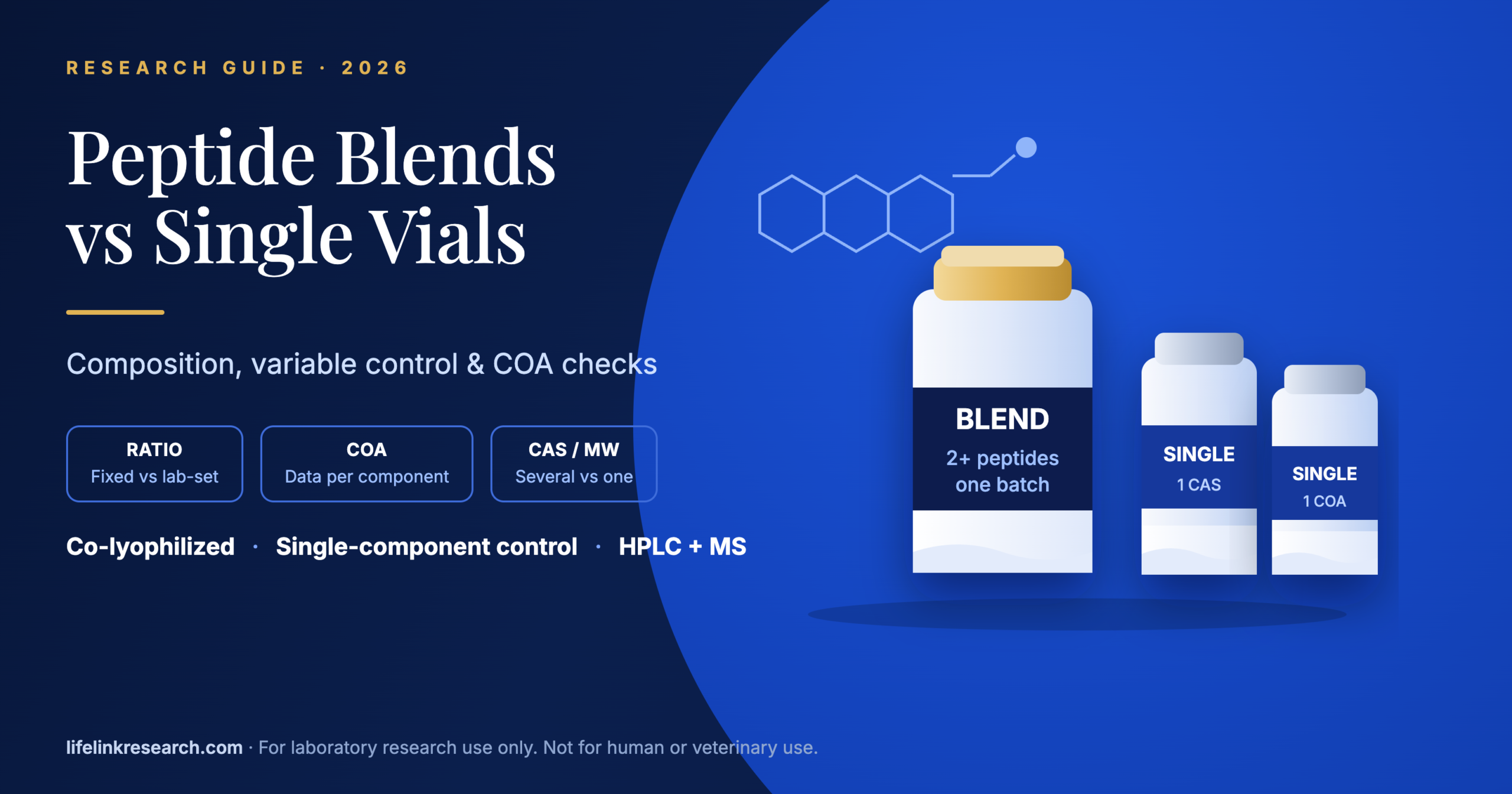
    Multi-Compound Blends: Reading a Blend COA

    A blend holds two or more peptides in one vial, so its COA has more to prove. Each component needs its own identity result, and ideally its own measured amount, because one matching mass can only speak for one molecule. Check that every compound on the label appears by name in the results, each with its own figure. Our KLOW stack COA guide walks through a four-component example field by field.

    FAQs

    Can peptide COAs be faked?

    Yes. Most fakes are genuine reports with an edited compound name, lot number or result, or one report reused across several batches. Altered reports rarely survive verification at the source. Entering the report’s key or ID on the lab’s portal, or emailing the lab, shows whether the results match what the lab actually issued.

    How do I look up a peptide COA by lot number?

    Find the lot or batch number on the vial label, then open the supplier’s COA page for that product and match the number exactly. Next, take the report ID or verification key from the COA to the lab’s own website. A peptide COA lookup is complete when both the supplier and the lab agree.

    What does 99% HPLC purity actually mean?

    It means 99% of the total peak area in the HPLC chromatogram belongs to the main peptide peak, measured at the lab’s detection wavelength. The remaining 1% is related impurities, such as shorter or modified sequences. The figure describes the tested sample only, and it says nothing about how much peptide the vial holds by weight.

    What’s the difference between purity and net peptide content?

    Purity, measured by HPLC, is the share of the peptide material that is the target sequence. Net peptide content asks a different question: how much of the weighed material is peptide at all once moisture and salts are subtracted. A sample can score high on purity and lower on net content, and a complete COA reports both.

    What is mass spectrometry used for on a COA?

    Mass spectrometry confirms identity. It measures the molecular weight of the compound in the sample and compares that observed value with the expected weight calculated from the peptide’s formula. A close match within the lab’s stated tolerance shows the main component is the labelled peptide, something an HPLC purity figure can’t establish alone.

    How recent should a peptide COA be?

    The COA should come from the same batch as the vial, so its date should fall around that batch’s production and before it went on sale. The age of the report counts for less than the match. A recent report for a different lot tells you less than an older one for the right lot.

    The Bottom Line

    A COA earns trust when it can be checked from the outside. The lot matches the label, the lab is real and accredited, the report verifies at the source, and the HPLC and mass-spec numbers fit the compound. Those four answers say more than any purity badge on a product photo. When the paperwork lines up, every Life Link product is supplied for laboratory research with its batch report one click from the product page.

    For Research Use Only. Not for use in diagnostic or therapeutic procedures.

    All products are supplied for in-vitro laboratory research by qualified professionals aged 21 and over. Read the full research use disclaimer.

    References

  • What Is TB-500? Thymosin Beta-4, Full-Length vs Fragment

    What Is TB-500? Thymosin Beta-4, Full-Length vs Fragment

    Quick Answer

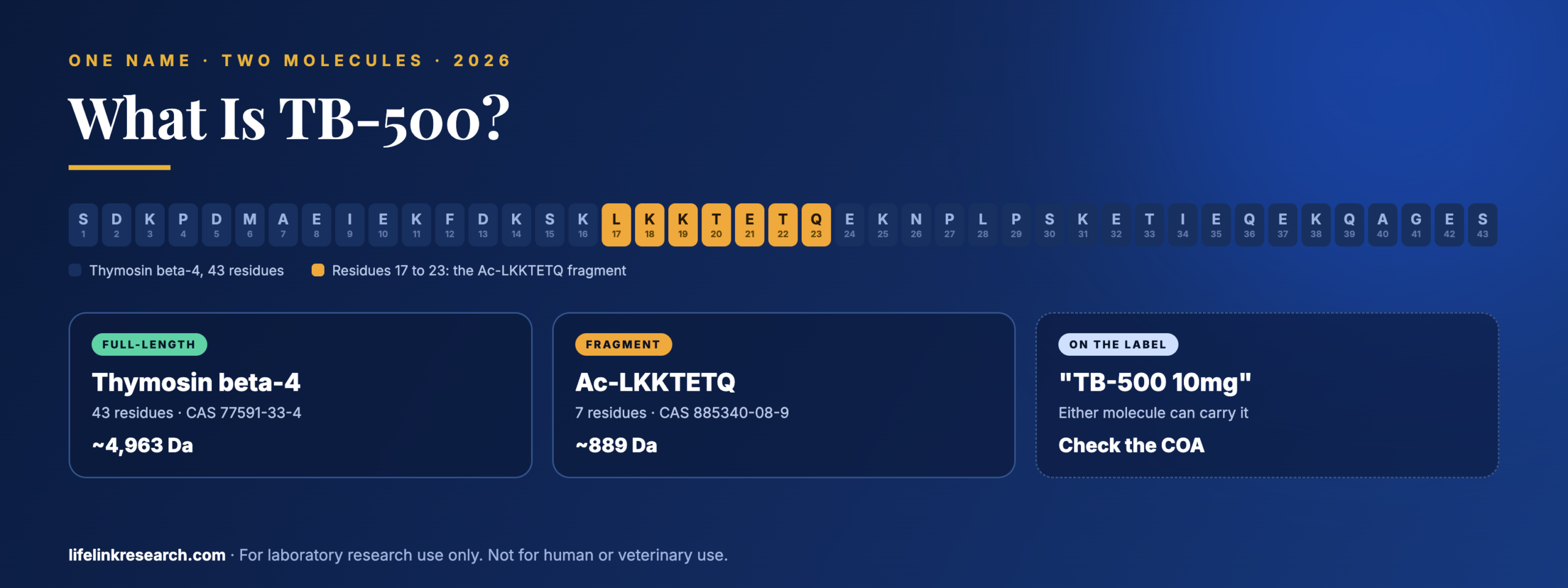
    TB-500 is a market name used for two different molecules. Some sellers mean full-length thymosin beta-4 (Tβ4), a 43-residue protein of about 4,963 Da. Others mean Ac-LKKTETQ, a 7-residue fragment of it of about 889 Da. A vial reading TB-500 10mg can hold either, and the identity result on its certificate is what resolves it.

    Key takeaways

    • TB-500 isn’t one molecule: it’s sold as full-length Tβ4 or a 7-residue fragment
    • Full-length Tβ4: 43 residues, about 4,963 Da, CAS 77591-33-4
    • The fragment: Ac-LKKTETQ, Tβ4 residues 17 to 23
    • The identity result on the COA tells you which one you have
    • Compare price per milligram of the same molecule

    The name and the vial often disagree. PubChem resolves TB-500 to a seven-residue peptide weighing about 889 daltons, while a great many vials sold under that name hold the whole 43-residue protein at roughly 4,963. Neither source is wrong. The label simply never belonged to one molecule, and that is the first thing to settle when asking what is TB-500.

    What follows covers both molecules, how the names came apart, where the published molecular weights disagree, and how a batch certificate resolves which one a given tb-500 peptide vial contains. Life Link’s own TB-500 10mg serves as the worked example.

    What Is TB-500?

    Quick answer: TB-500 is a vendor name, not a scientific one. It is used for either full-length thymosin beta-4, a naturally occurring 43-residue peptide, or for Ac-LKKTETQ, a synthetic 7-residue segment of it. Nothing has been reviewed by a regulator under that name, and the material is supplied purely for laboratory research.

    The designation came out of supplier catalogs, not from any journal or registry. Search the primary literature for thymosin beta-4 and the papers are plentiful; search it for “TB-500” and the term barely appears outside commercial contexts. That origin explains the looseness: a catalog label carries no obligation to specify a molecule, and two sellers can apply it to materials that differ fivefold in size.

    Two products ship under it today:

    • Full-length thymosin beta-4. The complete 43-residue peptide, acetylated at the N-terminus.
    • The Ac-LKKTETQ fragment. Seven residues taken from the middle of that sequence.

    Both are legitimate research materials, and they are not interchangeable.

    What Is Thymosin Beta-4?

    Quick answer: Thymosin beta-4 runs to 43 amino acids, carries an acetyl group on its N-terminus, and was first isolated from thymus tissue. It is one of the most studied members of the beta-thymosin family. In biochemical assays, it binds G-actin, the monomer form of actin, which is the property most research on it starts from.

    Structure

    The chain is catalogued in UniProt as P62328, where the mature 43-residue peptide runs:

    Ac-SDKPDMAEIEKFDKSKLKKTETQEKNPLPSKETIEQEKQAGES

    Three structural points govern how it behaves in a vial and on an instrument. It carries no cysteines, so there is no disulfide chemistry to preserve. Its single methionine at position 6 is the one residue that can pick up an extra 16 daltons through oxidation. And in solution the peptide is intrinsically disordered, taking on defined structure only when it binds actin, with no fixed fold of its own (Vitamins and Hormones, 2016, PMID 27450730).

    What the literature covers

    Published work on thymosin beta-4 is overwhelmingly cell-culture and animal-model research, with the G-actin binding interaction serving as the biochemical starting point for most of it. The beta-thymosin family is studied as a group, so papers frequently cover several members at once instead of Tβ4 alone. Thymosin alpha-1 is a separate 28-residue peptide despite the shared name; see what is thymosin alpha-1.

    TB-500 vs Thymosin Beta-4: What’s the Difference?

    Quick answer: Full-length thymosin beta-4 is the whole 43-residue molecule. The “TB-500” fragment is Ac-LKKTETQ, just residues 17 to 23, at about one-fifth of the mass. Sellers use both names loosely, so a vial labelled “TB-500” can contain either. They are different molecules and give different results on a COA.

    Full-length thymosin beta-4Ac-LKKTETQ fragment
    Residues437 (Tβ4 17 to 23)
    Molecular weightabout 4,963 Daabout 889 Da
    Monoisotopic mass4,960.49888.49
    CAS77591-33-4885340-08-9
    FormulaC212H350N56O78SC38H68N10O14
    PubChemCID 45382195CID 62707662
    OriginNaturally occurring peptide, made synthetically for researchSynthetic segment
    Molecules in 10mg2.0 µmol11.2 µmol, about 5.6× more

    Read the full sequence, and the fragment is easy to locate inside it: positions 17 to 23 spell LKKTETQ, falling just past the midpoint of the chain. That stretch is the part of the molecule most closely associated with actin binding, which is why it was isolated in the first place.

    Other fragments circulate too. Ac-SDKP is residues 1 to 4, taken from the opposite end of the same sequence, and it is sold under its own name, not as TB-500.

    One framing to retire: asking whether Tβ4 is “better” than TB-500 compares a molecule against a name. The useful question is which molecule a study calls for, and then which one the certificate confirms.

    What Is the Molecular Weight of Thymosin Beta-4?

    Quick answer: Full-length thymosin beta-4 has an average molecular weight of about 4,963 Da. Some sources list higher figures because they count a salt form or use a different calculation. The fragment Ac-LKKTETQ is about 889 Da. Always compare against the mass stated on the batch COA.

    PubChem puts the free-base average at 4,963 and the monoisotopic figure at 4,960.49. Figures above those appear often enough on secondary sites to cause confusion, and there are three ordinary reasons for the spread:

    • Average against monoisotopic. At this chain length, the two conventions differ by roughly 2.5 Da. Neither is wrong; they answer different questions, and a report should say which it used.
    • Free base against a salt. Research material is frequently supplied as an acetate. Every acetate counter-ion adds about 60 Da to the powder, and with nine lysines in the sequence there are plenty of sites to carry them, so a salt-form figure lands well above the free-base number.
    • Transcription errors. A figure copied between vendor pages without a source behind it tends to drift, which is why the arbiter is the batch document rather than a catalog.

    The same logic applies to the fragment, where 889 is the free-base average. Which questions each analytical method can answer is set out in HPLC vs mass spectrometry.

    How Do You Tell Which “TB-500” Is in Your Vial?

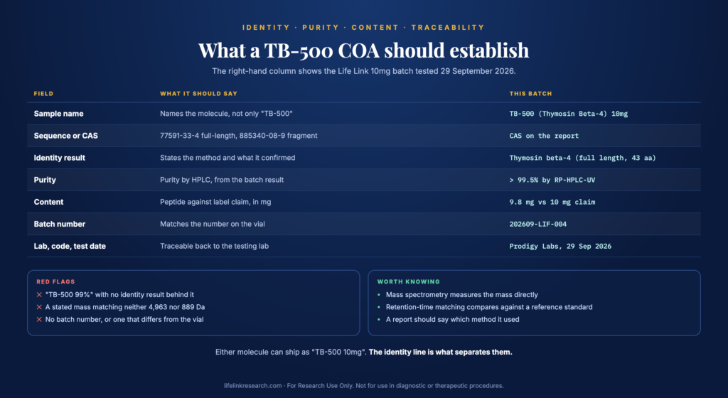
    Quick answer: Read the identity line on the COA. A measured mass near 4,963 Da means full-length thymosin beta-4; a mass near 889 Da means the Ac-LKKTETQ fragment. The report should also name the molecule, give its sequence or CAS, show HPLC purity, and carry a batch number matching your vial.

    What a TB-500 COA should establish: identity, purity, content and traceability

    What a TB-500 COA should establish: identity, purity, content and traceability

    Field on the COAWhat it should say
    Sample nameNames the molecule, not only “TB-500”
    Sequence or CAS77591-33-4 for full-length, 885340-08-9 for the fragment
    Identity resultThe method used, and what it confirmed
    PurityPurity by HPLC, read from the batch result
    ContentPeptide against label claim, in mg
    Batch numberMatches the number printed on the vial
    Lab, verification code, test dateMakes the report traceable back to the testing lab

    Three red flags cover most problems: a “TB-500 99%” line with no identity result behind it, a stated mass that matches neither molecule, and a missing batch number.

    How identity is actually established counts here. Mass spectrometry measures the molecule’s mass directly, which separates 4,963 from 889 beyond argument. Retention-time matching on HPLC is a different test: it compares the sample against a named reference standard and confirms they behave alike on the column. That is meaningful evidence when the reference standard is identified on the report, and it is worth knowing which of the two a certificate relied on.

    Life Link’s current 10mg batch, tested by Prodigy Labs on September 29, 2026 (report PL-COA-20260929-BF19-R4), records its identity as “Thymosin beta-4 (full length, 43 aa)”, confirmed by HPLC retention-time matching against a full-length acetate reference standard. Purity is reported above 99.5% by RP-HPLC-UV, and peptide content as 9.8 mg against a 10 mg label claim. So the material is the 43-residue protein. Checking any certificate against the issuing lab is covered in verify a COA is real.

    Why Does Price per mg Depend on Which Molecule?

    Quick answer: Ten milligrams of the fragment and ten milligrams of full-length thymosin beta-4 are very different products. The full-length protein is longer and more costly to make. Compare price per milligram only between listings that contain the same molecule, confirmed on their COAs.

    Synthesis cost scales with chain length. Each additional residue is another coupling step, another chance of a deletion sequence, and more purification work to remove what did not couple cleanly. A 43-residue chain is a substantially larger undertaking than a 7-residue one, and the catalog price follows.

    The molecule count runs the other way, which is what makes a naive comparison misleading. The same ten milligrams delivers about 2.0 µmol of the full protein or about 11.2 µmol of the fragment. A listing that looks cheap per milligram may simply hold the shorter molecule, so the only sound comparison is between listings whose certificates name the same compound.

    TB-500 in Blends and Kits

    Quick answer: TB-500 is a component of several multi-peptide products: with BPC-157 (often called “Wolverine”), with BPC-157 and GHK-Cu (GLOW), and with those plus KPV (KLOW). Each blend COA should list TB-500 separately, with its own mass result.

    Multi-component products raise the documentation bar rather than lowering it. A certificate covering two or four peptides has to resolve each one, which means a separate identity result per compound and a chromatogram showing a peak for each. A single combined purity figure describes none of them individually.

    For TB-500 specifically, the identity question carries straight into the blend: a kit listing “TB-500 10mg” inherits the same ambiguity as a standalone vial, and only the component-level result settles it. The BPC-157 + TB-500 kit keeps each compound in its own vial with its own certificate, which makes that check straightforward. The GHK-Cu, BPC-157 and TB-500 blend guide sets out the three-component composition.

    Is TB-500 Legal? FDA and WADA Status

    Quick answer: No FDA approval covers thymosin beta-4 or its fragments, and research-grade material goes to laboratories only. An FDA advisory committee took TB-500 up in July 2026 as a 503A compounding candidate, with no binding effect. Thymosin beta-4 also appears on the WADA Prohibited List.

    TB-500 appears on the first-day agenda of the July 23 to 24, 2026 advisory committee meeting, listed in free base and acetate forms. FDA stamps that page as current to August 6, 2026. A committee recommendation advises the agency and nothing more: a listing requires notice-and-comment rulemaking, and the 503A list concerns compounding pharmacies rather than reagent supply to laboratories. Dates and next steps are tracked in TB-500 and MOTS-c FDA status.

    In sport, the WADA Prohibited List effective January 1, 2026 names the compound under section S2, among growth factors, with the entry reading “Thymosin-ß4 and its derivatives e.g. TB-500”. The wording covers both the full protein and fragments taken from it, and the prohibition applies at all times.

    How Should TB-500 Be Stored?

    Quick answer: Freeze-dried vials belong at -20°C, sealed, dark, and dry. Warm a vial to ambient temperature first, then open it, which keeps condensation off the powder. Record the batch number against its certificate while the box is still to hand. These are storage conditions for the dry powder only.

    One sequence-specific note: with a single methionine at position 6 and no cysteines, oxidation at that one residue is the change most likely to show up on a later identity trace, as an extra 16 daltons. Keeping the vial sealed, cold, and dry is what holds the material close to the state its certificate describes.

    TB-500 FAQs

    Is TB-500 the same as thymosin beta-4?

    Sometimes. Some sellers use TB-500 for the complete 43-residue protein, others for the 7-residue Ac-LKKTETQ fragment taken from it. Because the same label covers both, the molecule named on the batch certificate is what settles it, not the product title.

    How many amino acids is TB-500?

    Either 43 or 7, depending on the molecule. Full-length thymosin beta-4 runs 43 residues with an acetylated N-terminus. The fragment sold under the same name is 7 residues, Ac-LKKTETQ, corresponding to positions 17 to 23 of the full sequence.

    What is Ac-LKKTETQ?

    It is a seven-residue peptide matching residues 17 to 23 of thymosin beta-4, acetylated at the N-terminus. PubChem lists it as CID 62707662, CAS 885340-08-9, formula C38H68N10O14, with an average mass near 889 Da. It is the region most associated with actin binding.

    What is the difference between TB-500 and BPC-157?

    They are unrelated molecules. BPC-157 is a synthetic 15-residue sequence of about 1,419 Da drawn from a gastric protein. TB-500 refers to thymosin beta-4 or its fragment, from an entirely different protein family. Our comparison of BPC-157 vs TB-500 covers both.

    Which TB-500 does Life Link sell?

    The full-length molecule. The current 10mg batch certificate, issued September 29, 2026, records identity as thymosin beta-4 at full length, 43 amino acids, with purity above 99.5% by HPLC and peptide content of 9.8 mg against the 10 mg label claim.

    Why do molecular weights differ between websites?

    Three reasons account for most of it: average mass against monoisotopic mass, which differ by roughly 2.5 Da here; free base against an acetate salt, where each counter-ion adds about 60 Da; and figures copied between pages without a primary source. The batch document settles it.

    ⏱ TB-500 in 30 seconds

    TB-500 is a catalog name covering two molecules. One is thymosin beta-4 itself: 43 residues, acetylated, about 4,963 Da, CAS 77591-33-4. The other is Ac-LKKTETQ, seven residues from positions 17 to 23 of that sequence, about 889 Da. Ten milligrams of the fragment contains roughly 5.6 times as many molecules. The identity result on the batch certificate is what tells the two apart.

    For Research Use Only. Not for use in diagnostic or therapeutic procedures. Read our full Research Use Disclaimer.

    What is described above is a laboratory reagent for qualified researchers aged 21 and over. The Terms and Conditions carry the conditions of sale in full.

    References

  • Wolverine Blend (BPC-157 + TB-500): Composition, COA & US Supply (2026)

    Wolverine Blend (BPC-157 + TB-500): Composition, COA & US Supply (2026)

    Quick Answer

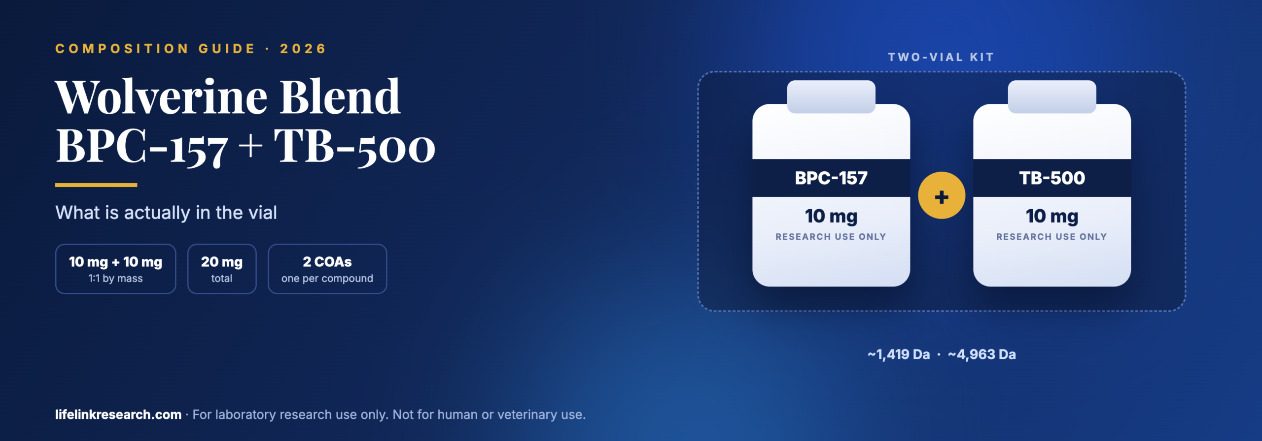
    “Wolverine” is the market nickname attached to a pairing of two research peptides, BPC-157 and TB-500, usually supplied at 10mg each (20mg in total, a 1:1 ratio). Suppliers ship it two ways: premixed in a single vial, or as a kit holding one vial of each. No regulator has approved it, and it is sold for laboratory research only.

    Key takeaways

    • Two compounds: BPC-157 (15 residues) and TB-500
    • Usually 10mg + 10mg, a 1:1 ratio
    • “TB-500” can mean the full protein or a 7-residue fragment, so check the label
    • A blend COA must show both compounds separately
    • Separate vials mean a separate COA for each

    Two vials can both read “TB-500 10mg” and hold molecules that differ more than fivefold in mass. One is thymosin beta-4, a 43-residue protein near 4,963 Da. The other is Ac-LKKTETQ, a seven-residue fragment near 889 Da. Same label, same milligrams, very different contents, and the wolverine blend is where that ambiguity turns up most often, because the name travels further than any specification sheet.

    What follows covers the composition of a wolverine peptide combination, the formats it ships in, how to read a two-component certificate, and what US supply looks like. Life Link’s own two-vial BPC-157 + TB-500 kit is used as the worked example throughout.

    What Is the Wolverine Blend?

    Quick answer: Wolverine is the catalog name given to BPC-157 and TB-500 when they are sold as a pair, most often at 10mg each. The nickname belongs to nobody, and no regulator has assessed anything bearing it. Labels vary between sellers, so the contents line and the batch certificate are the parts that actually describe the material.

    The nickname came from supplier catalogs and forum shorthand, not from any laboratory or registry. Nobody holds a trademark on it, which is exactly why two listings under the same name can hold different material.

    Three formats share the label:

    • Blend (one vial). Both peptides freeze-dried together in a single vial at a fixed ratio.
    • Kit (separate vials). Each peptide in its own vial, boxed together. Kit is the accurate word for this format, since nothing is actually combined.
    • Capsules. An oral format, outside the scope of this guide.

    Throughout the rest of this guide, the combination is called a BPC-157 + TB-500 blend, because that names what is in the vial.

    What Is in the Wolverine Blend?

    Quick answer: A standard Wolverine blend contains BPC-157 (10mg) and TB-500 (10mg), 20mg in total. BPC-157 is a synthetic 15-residue peptide; TB-500 is either full-length thymosin beta-4 or a short fragment of it, depending on the supplier.

    Every value below was read from PubChem and UniProt on October 25, 2026.

    ComponentAmountTypeSequence / sizeMolecular weightCAS
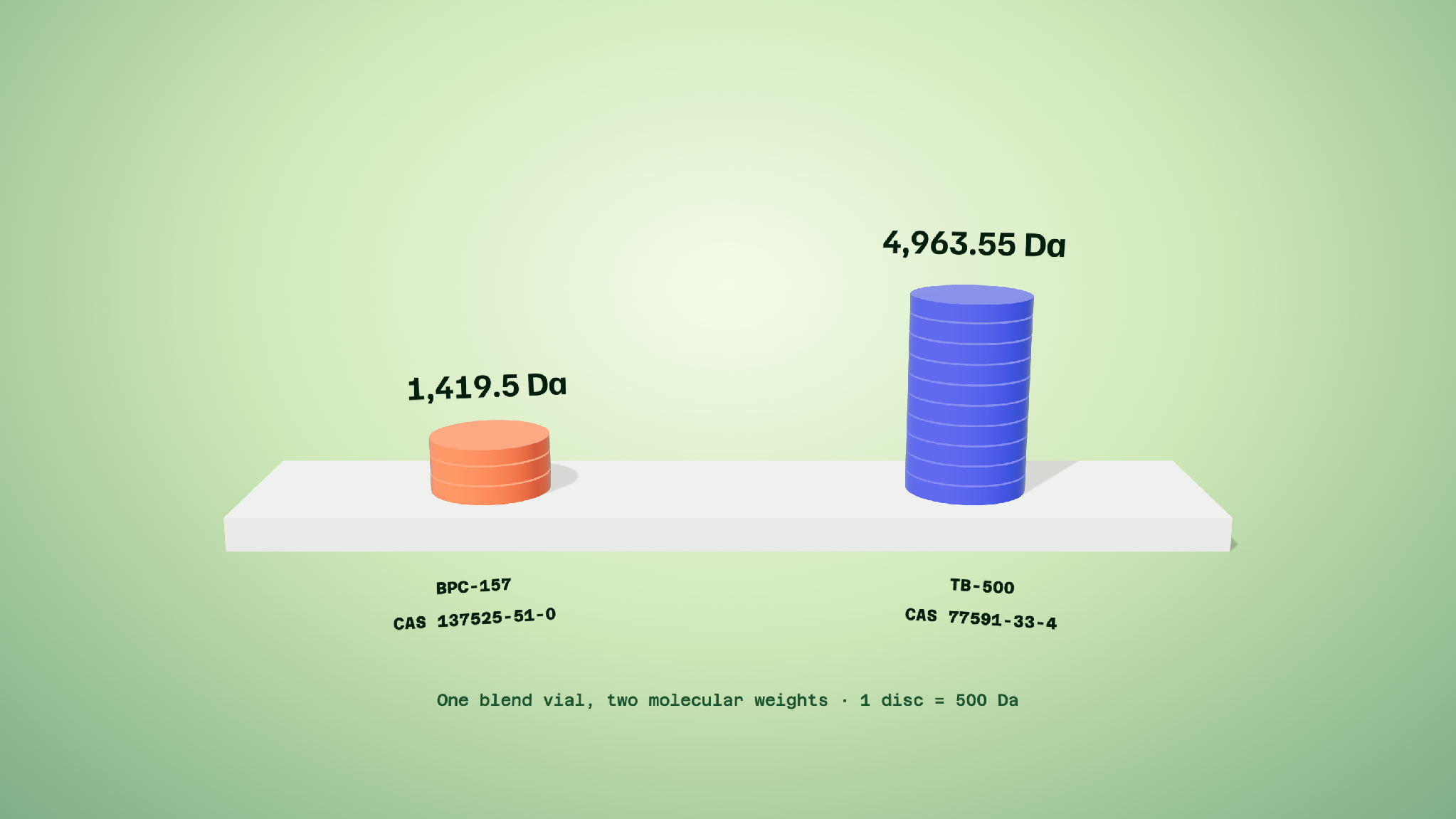
    BPC-15710 mgSynthetic 15-residue peptideGEPPPGKPADDAGLV~1,419.5 g/mol (CID 9941957)137525-51-0
    TB-500 (full-length)10 mgThymosin beta-4, 43 residues, N-acetylatedSDKPDMAEIEKFDKSKLKKTETQEKNPLPSKETIEQEKQAGES (UniProt P62328)~4,963 g/mol (CID 45382195)77591-33-4
    TB-500 (fragment)10 mgAc-LKKTETQ, a 7-residue fragmentLKKTETQ, acetylated~889 g/mol (CID 62707662)885340-08-9

    The BPC-157 row is settled: one sequence, one mass, one CAS number. The TB-500 rows are the reason this table has three entries instead of two.

    Which “TB-500” Is in Your Vial?

    Quick answer: Sellers use “TB-500” for two different molecules: full-length thymosin beta-4 (43 residues, about 4,963 Da) and a 7-residue fragment, Ac-LKKTETQ (about 889 Da). Both can be labelled “TB-500 10mg”. The COA’s measured mass tells you which one you bought.

    PubChem keeps them as separate records, and the names on those records are the clearest evidence that the field uses one label for two compounds. Search “TB-500” and you land on the 889 Da fragment. Search “thymosin beta-4” and you land on the 4,963 Da protein. Both are legitimate entries, and catalogs borrow freely from each.

    The arithmetic is where it lands:

    MoleculeMassMolecules per 10 mg (approximate)
    Thymosin beta-4, 43 residues~4,963 Da2.0 µmol
    Ac-LKKTETQ, 7 residues~889 Da11.2 µmol

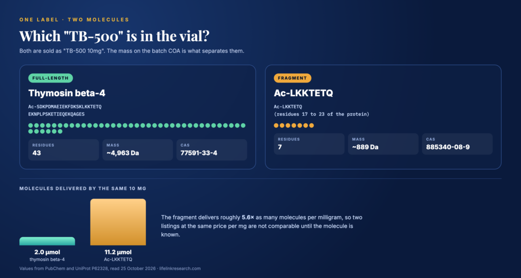
    Ten milligrams of the fragment delivers roughly 5.6 times as many molecules as ten milligrams of the full protein. Two listings at the same price per milligram are not comparable until you know which molecule each one holds.

    Two molecules sold as TB-500: thymosin beta-4 at 43 residues and the Ac-LKKTETQ fragment at 7 residues

    Two molecules sold as TB-500: thymosin beta-4 at 43 residues and the Ac-LKKTETQ fragment at 7 residues

    How to check: read the observed mass on the mass-spectrometry line of the batch COA. A result near 4,963 is the full protein; a result near 889 is the fragment. The product name on the label settles nothing on its own.

    Life Link’s TB-500 10mg is listed as full-length thymosin beta-4, CAS 77591-33-4, at 4,963.55 Da, and that CAS number and mass both correspond to the 43-residue protein. Confirm it against the mass on the current batch report before relying on it.

    Blended Vial vs Two-Vial Kit: Which Format?

    Quick answer: A blended vial holds both peptides freeze-dried together at a fixed 1:1 ratio, with one COA covering both. A two-vial kit keeps each peptide in its own vial with its own COA, so each can be checked separately and the ratio is set by the study, not the vial.

    FactorOne blended vialTwo-vial kit
    VerificationOne COA that must name both compoundsOne COA per compound
    RatioFixed at 1:1Set by the study design
    Stability questionsTwo peptides stored togetherEach stored apart
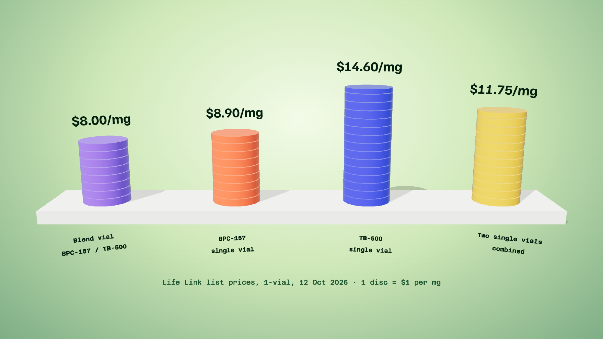
    Price per mgUsually lowerUsually higher

    The verification row is the one that separates the formats in practice. A single report covering a blended vial has to carry two identity results and two purity results, and plenty of them do not. With separate vials, each report is an ordinary single-compound certificate, which is far easier to check and far harder to fudge.

    The ratio row cuts the other way. A blended vial fixes the proportion at the point of manufacture, so a design calling for anything other than 1:1 cannot use it. Separate vials leave that choice open.

    Life Link stocks both: the BPC-157 + TB-500 10/10mg blended vial at $160, and the two-vial kit at $223.25. For the general case, blends vs single vials covers the trade-off across the catalog.

    What Should a Wolverine Blend COA Show?

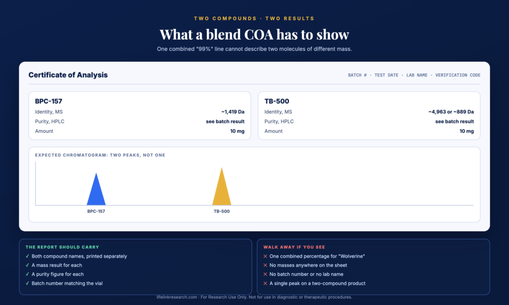
    Quick answer: A blend COA should list BPC-157 and TB-500 separately, each with its own identity result by mass spectrometry and its own purity result by HPLC. Expect two peaks on the chromatogram, not one. A single “Wolverine 99%” line doesn’t show that both compounds are present, or in what amount.

    What to look forWhy it belongs there
    Both compound names printed separatelyA combined name hides which component was measured
    Two mass results~1,419 for BPC-157, plus ~4,963 or ~889 for TB-500
    A purity figure for eachPurity by HPLC, read per compound, not as one number
    Two peaks on the chromatogramOne peak cannot represent two molecules of different mass
    Batch number matching the vialTies the document to the material in hand
    Lab name, verification code, and test dateLets the report be checked at source
    What a BPC-157 and TB-500 blend COA should show, with two peaks on the chromatogram

    What a BPC-157 and TB-500 blend COA should show, with two peaks on the chromatogram

    Red flags are the mirror image: one combined percentage, no masses, no batch number, no lab name.

    For a blended vial, the chromatogram is the quickest read. Two compounds at a 5.6-fold or 3.5-fold mass difference separate cleanly on a reverse-phase column, so a single peak on a two-component product is a question worth asking before purchase, not after. Our guides on how to verify a COA is real, and HPLC vs mass spectrometry cover the mechanics of each check.

    How Do BPC-157 and TB-500 Differ as Molecules?

    Quick answer: BPC-157 is a 15-residue synthetic sequence of about 1,419 Da. TB-500 is one of two things: thymosin beta-4 itself at 43 residues and roughly 4,963 Da, or the seven-residue piece cut from it. They differ in size, origin, and how they are characterised on a COA, which is why each needs its own result.

    BPC-157 runs fifteen residues, GEPPPGKPADDAGLV, and its sequence was taken from a larger protein identified in gastric juice. Nothing about it is extracted; the chain is assembled by solid-phase synthesis.

    Thymosin beta-4 is a naturally occurring 43-residue protein, catalogued in UniProt as P62328 and N-acetylated at its N-terminus. The LKKTETQ stretch inside it, residues 17 to 23, is the part the short fragment reproduces.

    That size difference has a consequence for the “1:1” shorthand. Equal milligrams are not equal molecules. Ten milligrams of BPC-157 is about 7.0 µmol, while ten milligrams of the 43-residue protein is about 2.0 µmol, so a 1:1 blend by mass is closer to 3.5:1 by molar amount. Against the short fragment the same 10mg pairing runs the other way, nearer 1:1.6. A protocol that specifies molar amounts needs the arithmetic done from the mass on the certificate.

    Published work on both compounds is mostly rodent and cell-culture studies. A fuller treatment of both molecules is in BPC-157 vs TB-500 compared.

    Is the Wolverine Blend Legal or FDA Approved?

    Quick answer: No FDA approval exists for BPC-157 or for TB-500, and nothing has ever been reviewed under the name “Wolverine”. In the US, both are supplied as research reagents for laboratory use. An FDA advisory committee took up both compounds in July 2026 for the 503A compounding list, and its recommendation carries no legal force.

    The FDA’s calendar entry for the July 23 to 24, 2026 advisory committee meeting names BPC-157 and TB-500 on the first day’s agenda, each listed in free base and acetate forms. The page is marked current as of August 6, 2026. That committee only advises. Anything reaching the 503A list has to clear notice-and-comment rulemaking first, and the list itself governs compounding pharmacies, not the supply of reagents to laboratories. BPC-157’s legal status tracks where that stands.

    In sport, both appear on the WADA Prohibited List effective January 1, 2026, in separate sections. The list places BPC-157 in S0, the non-approved substances class, while TB-500 falls under S2, where the entry reads “Thymosin-ß4 and its derivatives e.g. TB-500”. Both carry a ban that applies year-round.

    How Should the Wolverine Blend Be Stored?

    Quick answer: Keep the sealed freeze-dried vials in a -20°C freezer, shielded from light and kept dry. Bring a vial up to room temperature before breaking the seal, so moisture does not condense inside. Keep each kit vial labelled with its batch number next to its COA.

    With a two-vial kit, there is a small record-keeping point: the vials carry different batch numbers and arrive with different certificates. Log them separately on receipt, because once the box is discarded, matching a vial back to its report depends entirely on the label.

    Where to Buy the Wolverine Blend in the US

    Quick answer: Choose a US supplier that names both compounds on the label, publishes a separate identity and purity result for each, ships from US stock, and sells for research use only. Compare price per verified milligram, and check which TB-500 molecule each listing contains.

    Four checks separate a documented listing from a name on a box:

    • Both compounds named, with amounts, on the product page itself.
    • A batch COA for each compound, published and linked, with the lab named.
    • Which TB-500 molecule the listing contains, stated or evident from the mass.
    • US stock and research-use-only terms at checkout.

    Life Link supplies the combination as a two-vial kit: BPC-157 10mg and TB-500 10mg in separate vials, each with its own certificate, dispatched same day on orders placed before 2 PM PST and shipped UPS 2-Day Air from California. The blended 10/10mg vial is the alternative where a fixed 1:1 ratio suits the work. How to choose a US peptide supplier sets out the wider checklist.

    Wolverine Blend FAQs

    Why is it called “Wolverine”?

    The name spread through supplier catalogs and forum threads; it was never registered as a product. Since no business holds it and no agency has assessed anything carrying it, the contents line on the label and the batch COA describe a purchase far better than the nickname can.

    What ratio is the Wolverine blend?

    Usually 1:1 by mass: 10mg of BPC-157 and 10mg of TB-500, 20mg in total. That ratio is fixed in a blended vial. In a two-vial kit the amounts are the same, but each compound stays separate, so the proportion used is set by the study rather than by the manufacturer.

    Is Wolverine the same as GLOW or KLOW?

    No. The Wolverine combination holds two compounds. GLOW adds GHK-Cu to the same pair, making three, and KLOW adds both GHK-Cu and KPV, making four. The naming is catalog convention rather than chemistry, so check each listing’s composition. Adding GHK-Cu to the same two peptides gives the GLOW blend.

    Can I buy BPC-157 and TB-500 separately?

    Yes. Both are sold as single-compound vials, each with its own batch certificate, and buying them separately is what a two-vial kit amounts to. BPC-157 10mg is listed on its own at $89, with TB-500 10mg listed separately at $146.

    What is the difference between a blend and a kit?

    A blend is one vial holding both peptides freeze-dried together, documented by a single COA that has to cover both. A kit is two vials, each with its own COA. The blend fixes the ratio; the kit keeps the compounds and their paperwork separate.

    ⏱ Wolverine blend in 30 seconds

    Two research peptides sold under one nickname: BPC-157 at 10mg and TB-500 at 10mg, 20mg total in a 1:1 ratio. It ships either as a single blended vial or as a two-vial kit. The label “TB-500” covers two different molecules, near 4,963 Da and near 889 Da, so the mass on the batch COA identifies what is actually in the vial.

    For Research Use Only. Not for use in diagnostic or therapeutic procedures. Read our full Research Use Disclaimer.

    Everything described above is a laboratory reagent, sold only to qualified researchers aged 21 and over. Full sale conditions appear in the Terms and Conditions.

    References

  • What Is MOTS-c? Structure, Research Use & US Supply

    What Is MOTS-c? Structure, Research Use & US Supply

    Quick Answer

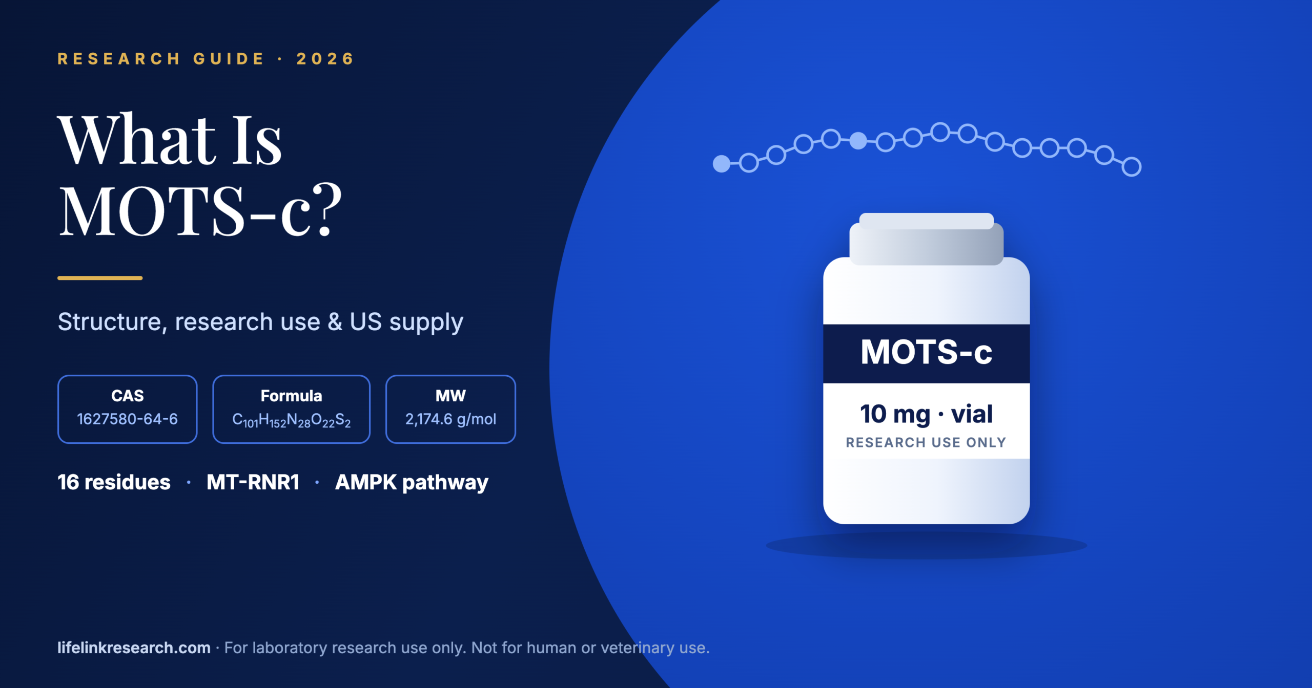
    MOTS-c is a 16-amino-acid peptide coded within a short open reading frame of the mitochondrial 12S rRNA gene (MT-RNR1). It was first described in 2015 and is classed as a mitochondrial-derived peptide. It’s studied in cell and animal models for AMPK pathway signaling and is supplied in the US as a lyophilized research reagent.

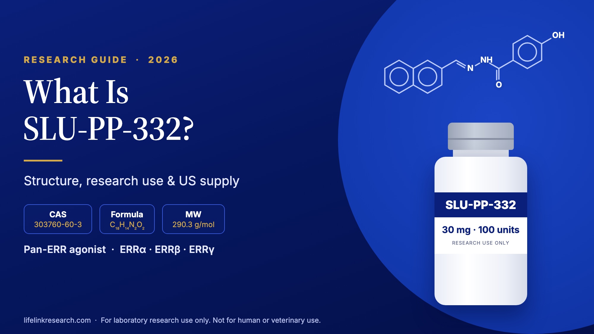
    Ribosomal RNA genes aren’t supposed to make proteins. Their RNA becomes part of the ribosome itself, so finding a peptide recipe tucked inside one surprised a lot of people. MOTS-c is that peptide, and it gives labs a short, fully defined molecule for studying how mitochondria send signals to the nucleus and cytoplasm. SLU-PP-332, a small-molecule ERR agonist, is another reference compound used in mitochondrial-signaling research. For a lab that wants a MOTS-c peptide with clean identity data, the structure is the place to start. New to peptide chemistry? Research peptides explained covers the basics.

    For Research Use Only. Not for use in diagnostic or therapeutic procedures.

    By Tyler Morgan · Scientific review by Kristen Doyle, PhD (biochemistry) · Updated October 18, 2026

    What Is MOTS-c?

    Quick answer: A 16-residue signaling peptide encoded in mitochondrial DNA, MOTS-c belongs to a small group called mitochondrial-derived peptides. A University of Southern California team, with Changhan Lee as first author and Pinchas Cohen as senior author, reported it in Cell Metabolism in 2015 (in vitro and mouse work). It’s made today by solid-phase synthesis.

    The 2015 paper (Cell Metabolism, 2015) described a short coding sequence (sORF) within the 12S rRNA gene and showed, in cell and mouse models, that its peptide acts on “the folate cycle and its tethered de novo purine biosynthesis, leading to AMPK activation.” That pathway language is still how the field describes MOTS-c today. Our research library covers related peptides in the same style. For example, the Epithalon (AEDG) structure and COA guide covers a four-residue synthetic peptide.

    What does MOTS-c stand for?

    The name is a compressed description of where the peptide comes from: Mitochondrial Open reading frame of the Twelve S rRNA, type c. UniProt lists the same expansion, with the gene name MT-RNR1, and a 2023 review in Antioxidants spells it out in full as “Mitochondrial open reading frame of the 12S rRNA type-c” (review, 2023).

    Mitochondrial-derived peptides: humanin, SHLPs and MOTS-c

    MOTS-c has two better-known relatives. Together they form a family defined by origin, not by shared sequence.

    PeptideLengthGene regionFirst reported
    Humanin24 residues16S rRNA (MT-RNR2)2001
    SHLP1 to SHLP620 to 38 residues16S rRNA (MT-RNR2)2016 (Cobb et al., in vitro and animal)
    MOTS-c16 residues12S rRNA (MT-RNR1)2015

    Of the three, only MOTS-c maps to the 12S region, and it’s also the shortest.

    What Is the Structure of MOTS-c?

    Quick answer: MOTS-c is an unbranched chain of 16 residues with the sequence MRWQEMGYIFYPRKLR. Its formula is C101H152N28O22S2, with an average molecular weight near 2174.6 Da. It carries two methionines, one tryptophan, two tyrosines and a run of basic residues near the C-terminus.

    Each row was cross-checked on October 18, 2026 with PubChem CID 146675088, UniProt A0A0C5B5G6 and the FDA GSRS record.

    FieldValue
    Sequence (one-letter)MRWQEMGYIFYPRKLR
    Sequence (three-letter)Met-Arg-Trp-Gln-Glu-Met-Gly-Tyr-Ile-Phe-Tyr-Pro-Arg-Lys-Leu-Arg
    Length16 amino acids
    Molecular formulaC101H152N28O22S2
    Average molecular weight2174.6 Da
    Monoisotopic mass2173.1077 Da
    UniProt IDA0A0C5B5G6 (MOTSC_HUMAN)
    CAS1627580-64-6
    FDA UNIIA5CV6JFB78
    Gene originMT-RNR1 (12S rRNA), mitochondrial DNA
    FormLyophilized powder

    Reading the sequence

    Walk along the chain and three features jump out:

    • Two methionines (M1 and M6). Both carry sulfur, the “S2” in the formula, and both can oxidize. Each oxidation adds 16 Da, a change a mass spectrum picks up easily.
    • An aromatic core (W3, Y8, F10, Y11). These residues give MOTS-c strong UV absorbance at 280 nm, which helps on an HPLC trace.
    • A basic tail (R13, K14, R16). Add R2 and the N-terminal amine, and the peptide carries about five positive charges against one acidic glutamate (E5) and the C-terminal acid. At neutral pH that leaves a net charge near +3.

    That charge profile explains why MOTS-c shows up mostly as 3+ and 4+ ions in electrospray mass spectrometry, a point the COA section comes back to.

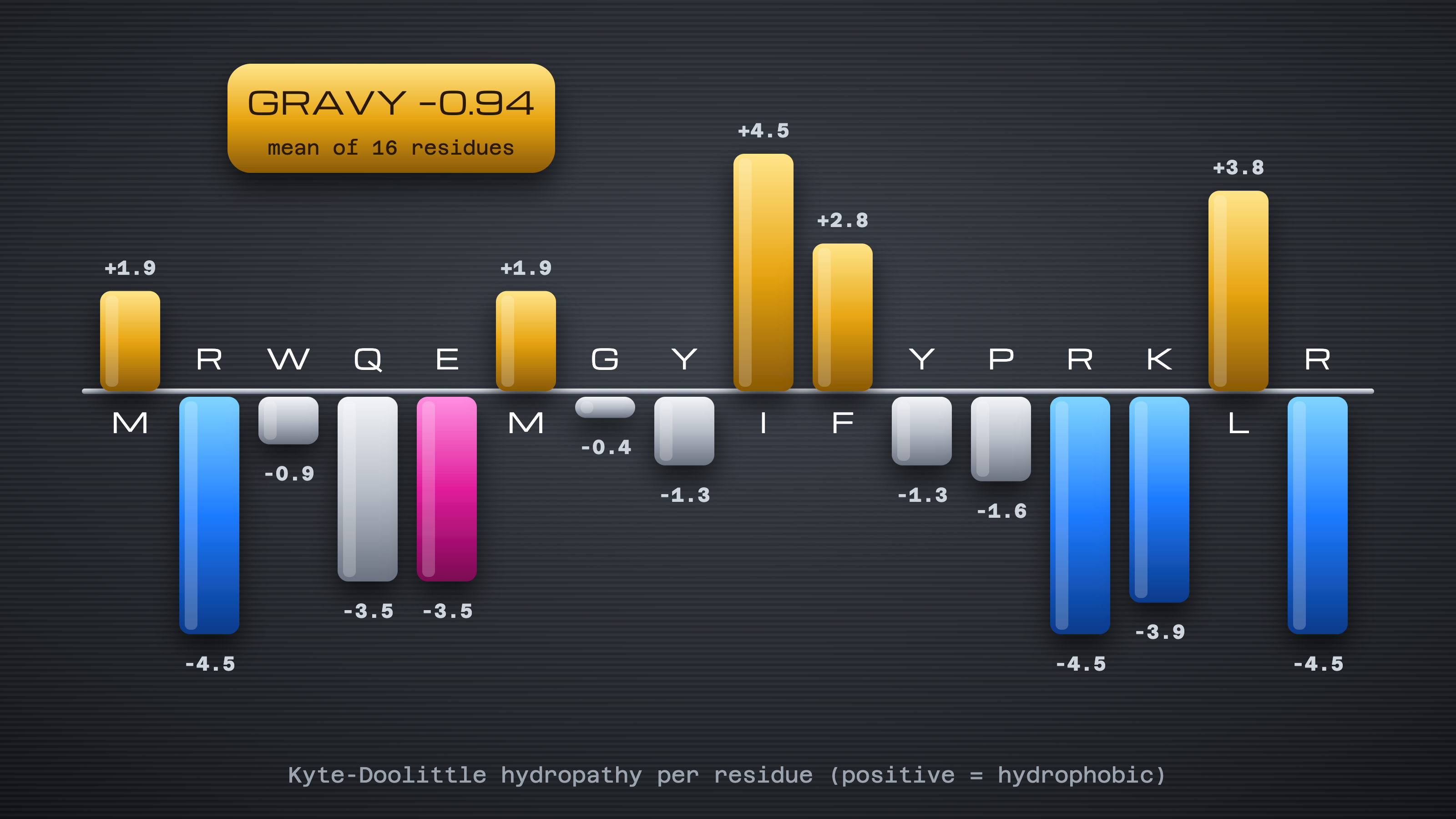
    Kyte-Doolittle hydropathy bars for each MOTS-c residue, with a hydrophobic Ile-Phe core, a charged Arg-Lys tail and a GRAVY score of -0.94

    Each MOTS-c residue, with a hydrophobic Ile-Phe core, a charged Arg-Lys tail and a GRAVY score of -0.94

    Gene origin: a peptide encoded in mitochondrial DNA

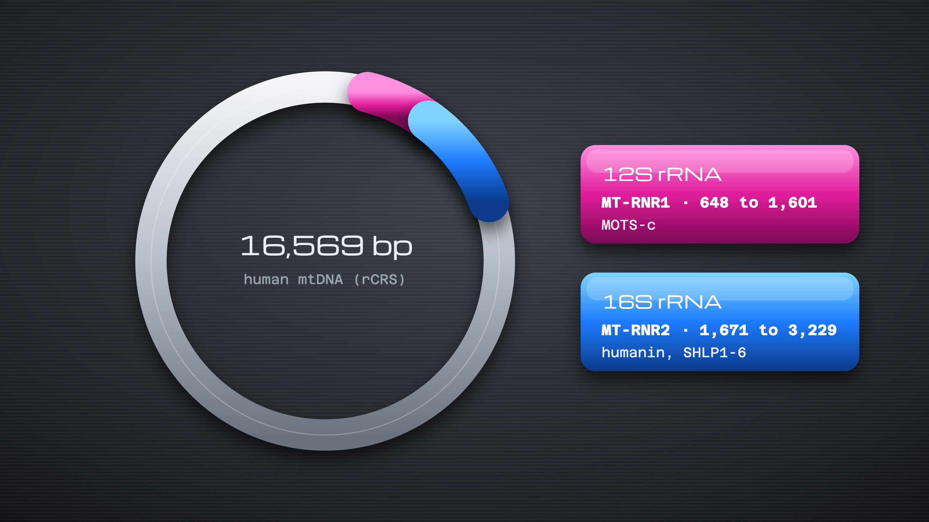
    Human mitochondria carry their own small circular genome of about 16.6 kb. Most of it encodes respiratory-chain subunits, transfer RNAs and the two ribosomal RNAs. MOTS-c’s coding stretch overlaps the 12S rRNA gene, so the same stretch of DNA serves two purposes. Kim and colleagues later described this arrangement as evidence “that mitonuclear communication is genetically integrated” (Cell Metabolism, 2018).

    Anodized ring map of the 16,569 bp human mitochondrial genome highlighting the 12S rRNA gene (MT-RNR1, 648 to 1,601), home of MOTS-c, and the 16S rRNA gene (MT-RNR2, 1,671 to 3,229), home of humanin and SHLP1-6

    16,569 bp human mitochondrial genome highlighting the 12S rRNA gene (MT-RNR1, 648 to 1,601), home of MOTS-c, and the 16S rRNA gene (MT-RNR2, 1,671 to 3,229), home of humanin and SHLP1-6

    Handling properties for the lab

    • Storage: −20 °C, desiccated, in the sealed vial.
    • Solubility: soluble in water for assay preparation.
    • Watch points: methionine oxidation and moisture uptake are the main ways a dry sample drifts from its original COA.

    How Is MOTS-c Used in Research?

    Quick answer: MOTS-c is studied in in vitro and rodent models as a tool for AMPK pathway signaling, folate-cycle and AICAR assays, and nuclear translocation studies. Human data are limited to observational measurements and registered studies, so each finding needs its model type attached before it’s compared with anything else.

    AMPK and folate-cycle signaling

    The 2015 paper placed MOTS-c upstream of AMPK through the folate cycle. In that model (cell lines and mice), MOTS-c slowed folate-cycle flux, which let AICAR, an intermediate in de novo purine synthesis, build up. AICAR is a known AMPK activator, so a typical assay pairs AICAR measurement with AMPK phosphorylation at Thr172 by western blot. Many labs add a second read-out downstream, phosphorylation of the AMPK substrate acetyl-CoA carboxylase (ACC) at Ser79, to confirm the pathway is engaged and not just the kinase itself.

    Nuclear translocation and gene-expression studies

    Three years later, the same lab showed in cultured cells that MOTS-c moves into the nucleus under glucose restriction, in an AMPK-dependent manner. There it regulated genes carrying antioxidant response elements (ARE) and interacted with the transcription factor NRF2 (in vitro, Cell Metabolism, 2018). These experiments rely on cell fractionation, imaging of tagged peptide and transcript profiling.

    Study types at a glance

    ModelWhat is measuredSource
    Cell lines (in vitro)Folate-cycle metabolites, AICAR levels, AMPK phosphorylation by western blotCell Metabolism, 2015
    Cell lines (in vitro)Nuclear vs cytoplasmic peptide, ARE-gene transcript levels, NRF2 bindingCell Metabolism, 2018
    RodentPathway markers in tissue samples by western blotCell Metabolism, 2015
    Human, observationalCirculating peptide quantified by ELISAClinical Endocrinology, 2019; Experimental Physiology, 2026
    Human, registered studyNCT07505745: Phase 2, interventional, recruiting since February 2026ClinicalTrials.gov, checked October 18, 2026

    What the research does not establish

    No regulator has approved MOTS-c for any human use. Results from cell lines and rodents describe how a pathway behaves in that model, and they don’t carry over to people by default. The single registered human study is still recruiting, with no posted results as of October 2026. Observational studies, for their part, measure how much MOTS-c peptide circulates; they don’t test what it does. Keeping those categories apart is the simplest way to read the literature accurately.

    Is MOTS-c FDA Approved?

    Quick answer: Not as of October 18, 2026. There’s no FDA-approved MOTS-c product, and openFDA’s Drugs@FDA database returns no record for it. US suppliers offer it only as a lab research reagent. A compounding vote in July 2026 left its approval status unchanged.

    Each line below is dated and sourced:

    • Compounding review. On July 23 and 24, 2026, MOTS-c came before FDA’s Pharmacy Compounding Advisory Committee as a 503A Bulks List candidate, and the panel voted 7 for, 5 against, with 2 abstentions, to recommend it (source: FDA meeting page, July 2026).
    • Status after the vote. The committee advises FDA. A listing still needs notice-and-comment rulemaking, so MOTS-c’s 503A category is unchanged as of October 2026 (source: Federal Register, April 16, 2026).
    • WADA. The 2026 Prohibited List, effective January 1, 2026, names MOTS-c under S4 (hormone and metabolic modulators) as an example of an AMPK activator, alongside AICAR (source: WADA Prohibited List, 2026).

    For the full timeline and what happens next, read MOTS-c and TB-500 FDA status (2026), and for the other six peptides on that agenda, see FDA’s July 2026 peptide review.

    How Is MOTS-c Supplied for Research in the US?

    Quick answer: Research MOTS-c in the US typically comes as a 10 mg lyophilized vial with a lot number that links to its batch COA. Life Link ships from California and sells only under research-use-only terms to researchers aged 21 or older, so each order arrives documented and traceable to a tested lot.

    What arrives and how to store it on receipt

    The MOTS-c 10 mg research peptide arrives as a sealed vial in plain outer packaging. On receipt:

    1. Check the seal and compare the vial’s lot number with its COA.
    2. Move it to −20 °C, dry and dark.
    3. Log the lot, receipt date and freezer location.

    The dry, freeze-dried format is deliberate. With two methionines in the sequence, MOTS-c is more exposed to oxidation than many short peptides, and removing water slows that chemistry. A sealed, cold, dark vial keeps the sample close to the state its COA describes.

    Ordering terms and US shipping

    Same-day dispatch applies to orders in by 2 PM PST, sent UPS 2-Day Air out of California, with tracking by email. Every sale carries research-use-only terms, and buyers confirm they’re 21 or older. Carrier options and timings are set out in the US shipping policy.

    What Should a MOTS-c COA Show?

    Quick answer: A MOTS-c COA should show HPLC purity with its chromatogram, a mass-spectrometry identity result matching about 2174.6 Da, net peptide content, and the lot number, test date and testing lab. Purity by HPLC, see the batch result. Everything on the report should trace to the lot on your vial.

    TestWhat it confirmsWhat to check
    HPLC purityShare of UV signal in the main peakA chromatogram is attached, not just a number
    MS identityThe molecule is MOTS-cCharge-state ions near 1088.06 (2+), 725.71 (3+) and 544.28 (4+), or a deconvoluted mass near 2173.1 Da
    Net peptide contentPeptide vs counter-ions and waterStated as a percentage; with five basic sites, counter-ions can make up a large share of the powder
    Counter-ionSalt form (acetate or TFA)Named on the report; TFA often carries over from synthesis unless the batch was exchanged to acetate
    Lot, date, labTraceabilityMatches your vial; lab named; date close to manufacture

    The charge-state values above are calculated from PubChem’s monoisotopic mass. A peak 16 Da heavier on any of them points to oxidized methionine. Every Life Link lot is listed in the batch COA library.

    Red flags on a peptide COA

    • The lot number is missing or differs from the vial.
    • Purity appears as a bare number.
    • The reported mass fits neither 2174.6 Da nor a known charge state.
    • One report is reused across several lots.
    • No lab name or test date appears.

    Our guide on how to verify a peptide COA walks through each check.

    MOTS-c FAQs

    How many amino acids are in MOTS-c?

    MOTS-c has 16 amino acids: MRWQEMGYIFYPRKLR. That’s short enough to make by solid-phase synthesis in one run, and long enough that electrospray tends to show it carrying several charges at once. UniProt lists it as MOTSC_HUMAN (A0A0C5B5G6), encoded within the mitochondrial MT-RNR1 gene.

    Who discovered MOTS-c?

    A University of Southern California group led by Changhan Lee, with senior author Pinchas Cohen, reported MOTS-c in Cell Metabolism in March 2015. Their work combined cell-line experiments and mouse models. Lee’s lab followed up in 2018 with the nuclear translocation study, also in Cell Metabolism and also in cultured cells.

    Is MOTS-c on the WADA Prohibited List?

    Yes. WADA’s 2026 Prohibited List, in force since January 1, 2026, names MOTS-c under section S4, hormone and metabolic modulators, as an example of an activator of AMP-activated protein kinase (AMPK). It appears there alongside AICAR.

    Is MOTS-c the same as humanin?

    No. Both are mitochondrial-derived peptides, but they’re different molecules from different genes. Humanin has 24 residues and comes from the 16S rRNA region (MT-RNR2). MOTS-c is 16 residues long and traces to the 12S region (MT-RNR1). Their sequences don’t overlap, so their mass-spectrometry identities are entirely distinct.

    Does Life Link publish a COA for every MOTS-c batch?

    Yes. Each lot goes to Janoshik Analytical or a US-based lab for two checks, LC-MS for identity and RP-HPLC for purity, and its report carries the batch number and test date. A wider panel (heavy metals, endotoxin, sterility) runs on rotation. See our testing process.

    Closing Notes

    MOTS-c is a compact, well-mapped peptide: 16 residues, one mitochondrial gene and a pathway literature built mostly on cell and rodent models. For lab work, the identity data on its COA count for more than any label. Product details and the current batch report are on the MOTS-c product page linked above.

    Lab use only. For Research Use Only. Not for use in diagnostic or therapeutic procedures. MOTS-c from Life Link Research is sold to professionals as a laboratory reagent. FDA has not reviewed or approved it, and this article offers no medical advice. See our Terms and Conditions for the conditions of sale. Read our full Research Use Disclaimer. Last updated October 18, 2026.

    References

    Access date for every source: October 18, 2026.

    • Lee C, et al. Cell Metabolism. 2015;21(3):443-454. In vitro and mouse study. doi:10.1016/j.cmet.2015.02.009
    • Kim KH, et al. The mitochondrial-encoded peptide MOTS-c translocates to the nucleus to regulate nuclear gene expression in response to metabolic stress. Cell Metabolism. 2018;28(3):516-524. In vitro. doi:10.1016/j.cmet.2018.06.008
    • Cobb LJ, et al. 2016;8(4):796-809. In vitro and animal study. doi:10.18632/aging.100943
    • PubChem CID 146675088; UniProt A0A0C5B5G6; FDA GSRS UNII A5CV6JFB78
    • ClinicalTrials.gov NCT07505745
    • FDA Pharmacy Compounding Advisory Committee, July 23 and 24, 2026
    • WADA Prohibited List, 2026 edition
  • What Is DSIP? Structure, Research Use & US Supply

    What Is DSIP? Structure, Research Use & US Supply

    DSIP is a synthetic nine-residue peptide, WAGGDASGE, listed under the international name Emideltide. On July 24, 2026, it drew the narrowest vote of an FDA advisory meeting on compounding peptides, and the agency’s briefing pointed less at biology than at chemistry: how well the molecule is characterized and which impurities might travel with it.

    Those are lab questions, and this page answers them in the same spirit. You’ll find the sequence and identifiers, the kinds of assays DSIP turns up in, how to read its certificate of analysis, how sealed vials are stored, its US status and what to ask a supplier. More reference pages are in our research library.

    For Research Use Only. Not for use in diagnostic or therapeutic procedures.

    What Is DSIP?

    Quick answer: DSIP is a linear nonapeptide with the sequence WAGGDASGE, also listed as Emideltide. It was first isolated from rabbit cerebral venous blood in the 1970s by a Basel group. Today it’s made by chemical synthesis and supplied as a lyophilized research peptide for analytical and in vitro work, under research-use-only rules.

    The Basel lab of Guido Schoenenberger and Marcel Monnier spent years on the fraction before its sequence appeared. The final isolation and characterization were published in 1977, in Pflügers Archiv (animal tissue source, 1977) and in PNAS (1977). The Pflügers Archiv paper was part X of a series, which explains why some sites quote earlier dates for the “discovery.” The 1977 papers are the ones that put the nine residues on record.

    Two things changed after that:

    • Synthesis replaced extraction. Once the sequence was known, chemists could build it residue by residue on solid-phase resin. The same group compared native and synthetic material in Experientia that April (1977). Every DSIP peptide vial sold for research today is synthetic.
    • The molecule got an INN. “Emideltide” is the international nonproprietary name, and it’s the term FDA uses in its compounding documents. Same peptide, two names, so search both when you check a source.

    A 2006 mini-review by Vladimir Kovalzon and Tatyana Strekalova in the Journal of Neurochemistry noted that “DSIP structure is different from any other known representative of the various peptide families” (review, 2006). That uniqueness is part of why analysts treat it as a clean test case: there’s no close natural cousin to confuse it with.

    Where the name comes from

    DSIP’s name is a historical label. The Basel team named the peptide after the experimental system in which they first tracked the fraction, and the label stuck long before anyone could explain what the molecule does. It’s nomenclature, the way “secretin” or “substance P” reflect the labs that coined them. It isn’t a confirmed description of a property.

    That’s also why you’ll see the full phrase, delta sleep inducing peptide, in older indexes and catalogs, while newer regulatory papers default to Emideltide. Kovalzon and Strekalova’s review points out that no gene, precursor protein or receptor for DSIP had been isolated as of 2006, so the name still points back to its origin rather than to a mapped pathway. For anyone searching the literature, use all three terms: DSIP, delta sleep-inducing peptide and Emideltide.

    What Is the Structure of DSIP?

    Quick answer: DSIP is a 9-amino-acid linear peptide: Trp-Ala-Gly-Gly-Asp-Ala-Ser-Gly-Glu. Its formula is C35H48N10O15, its molecular weight is about 848.8 g/mol, its CAS number is 62568-57-4 and its PubChem CID is 68816. It ends in an unblocked amine at one terminus and an unblocked carboxylic acid at the other, and it’s supplied as a white lyophilized powder.

    Amino acid sequence

    Three glycines and two alanines give DSIP a small, flexible backbone. Tryptophan at position 1 is the only aromatic residue and the only one with strong absorbance at 280 nm, which turns out to be useful on an HPLC trace. Epithalon (Ala-Glu-Asp-Gly) is another short synthetic peptide built from glycine and acidic residues; see the Epithalon structure and COA guide.

    PositionThree-letterOne-letterSide-chain class
    1TrpWAromatic, hydrophobic
    2AlaASmall, nonpolar
    3GlyGNo side chain
    4GlyGNo side chain
    5AspDAcidic
    6AlaASmall, nonpolar
    7SerSPolar, hydroxyl
    8GlyGNo side chain
    9GluEAcidic

    Count the charges and a pattern appears. Asp5, Glu9 and the C-terminal carboxyl are all acidic, while the N-terminal amine is the only basic group. At neutral pH, DSIP works out to a net charge near −2, which is unusual among research peptides. On a reversed-phase column it elutes early, and in mass spectrometry it ionizes well in negative mode, a detail the COA section returns to.

    Physicochemical properties

    All values below were checked against PubChem CID 68816 and FDA GSRS on October 18, 2026.

    PropertyValue
    Molecular formulaC35H48N10O15
    Average molecular weight848.8 g/mol
    Monoisotopic mass848.3301 Da
    CAS number62568-57-4
    PubChem CID68816
    FDA UNIIYN28Z5YZ73 (Emideltide)
    Computed XLogP−7.1 (strongly hydrophilic)
    AppearanceWhite lyophilized powder
    SolubilitySoluble in water for assay preparation

    Purity is left out of this table on purpose: it belongs to a batch, and each lot gets its own figure. Our explainer on peptide purity 98% vs 99% shows what that last percentage point represents.

    Phosphorylated and analog forms in the literature

    DSIP has a phosphorylated relative. Adding a phosphate to Ser7 gives P-DSIP, which raises the average mass by about 80 Da to roughly 928.8 g/mol. It has been prepared synthetically and appears in recent animal research (Life Sciences, 2018).

    Researchers have also built structural analogs with substituted residues. The 2006 review notes a naturally occurring decapeptide that matches DSIP at five of nine positions. GSRS separately registers N-acetyl Emideltide (UNII CL985USD2K), a reminder to confirm which form a document describes.

    How Is DSIP Used in Research?

    Quick answer: In labs, DSIP is used as a reference standard in HPLC and LC-MS method development, in peptide stability and enzymatic degradation assays, and in immunoassays for DSIP-like immunoreactivity in tissue extracts. Researchers should note which findings come from in vitro work, which from animal models, and which from older human literature.

    A short, well-defined, acidic peptide with one UV-active residue is handy for analytical chemists. DSIP works as a test analyte when tuning gradients, checking negative-mode mass spectrometry, or comparing column chemistries for polar peptides.

    The published literature, meanwhile, falls into three study types. The table lists what kind of work exists in each, not what it found.

    In vitroAnimalHuman
    Degradation and aggregation studies in plasma and serum (Peptides, 1987)Original isolation from rabbit tissue (1977)Small clinical reports from the 1980s, such as a pilot study in European Neurology (1984)
    Analytical method work: HPLC, LC-MS, peptide mappingImmunohistochemical mapping of DSIP-like immunoreactivity in tissuesMeasurement of DSIP-like immunoreactivity in plasma samples
    Antibody and immunoassay developmentComparisons of native vs synthetic material (1977)Few studies, mostly before 2000

    One caution applies to immunoassays. In 1993, a team characterized a 77-residue protein from porcine brain that an anti-DSIP antiserum also recognized (European Journal of Biochemistry, 1993). “DSIP-like immunoreactivity” therefore doesn’t always mean DSIP itself, and mass spectrometry is the tiebreaker.

    What’s still missing is a target. As Kovalzon and Strekalova’s review describes, no specific DSIP receptor has been isolated. Receptor-binding assays, which anchor work on most peptides, have no confirmed partner to run against here. That’s one reason so much DSIP literature is analytical or descriptive: identity, stability, distribution of immunoreactivity. For a lab, it also means every experiment leans hard on knowing exactly what’s in the vial.

    How Do You Read a DSIP COA?

    Quick answer: A DSIP COA should show the lot number, HPLC purity with a chromatogram, and LC-MS identity matching about 848.8 Da. It should also give appearance, test date and the testing lab. Purity is reported per batch, so check the result for your lot rather than a headline figure. Endotoxin or heavy-metal panels appear only if they were run.

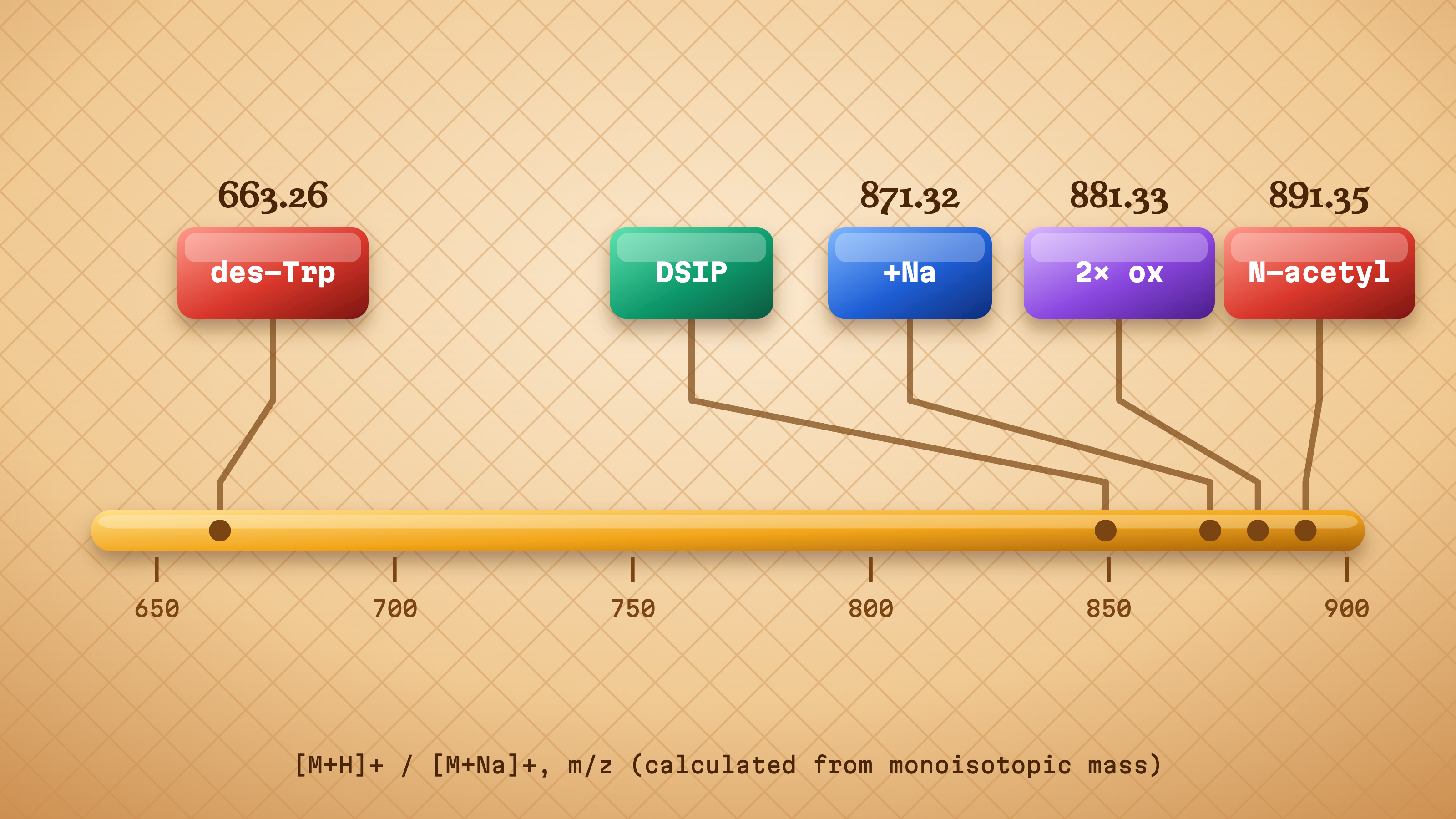
    These are the ions to look for, calculated from the monoisotopic mass of 848.3301 Da:

    IonExpected m/zWhen you’ll see it
    [M+H]+849.34Positive-mode ESI, the usual default
    [M+2H]2+425.17Positive mode, smaller share for an acidic peptide
    [M−H]−847.32Negative mode, which suits DSIP’s acidic residues
    [M+O+H]+865.33Trp oxidation product; should be absent or a trace

    For a field-by-field walkthrough of any certificate, see how to read a peptide COA.

    HPLC purity vs MS identity

    Each test covers a different blind spot. HPLC pulls the sample apart on a column and measures what share of the UV signal lands in the main peak. LC-MS weighs that peak to show it really is DSIP.

    DSIP adds a twist. Its tryptophan absorbs at 280 nm, while every peptide bond absorbs at 214 nm. A COA that shows both wavelengths can flag impurities that lack the Trp residue, such as truncated sequences missing position 1. HPLC and mass spectrometry trade off differently: one quantifies, the other identifies.

    Mass ruler of calculated DSIP-related ions: des-Trp 663.26, sodium adduct 871.32, double oxidation 881.33 and N-acetyl 891.35, beside the protonated DSIP ion

    Mass ruler of calculated DSIP-related ions: des-Trp 663.26, sodium adduct 871.32, double oxidation 881.33 and N-acetyl 891.35, beside the protonated DSIP ion

    Red flags on a peptide COA

    Run down this list before filing any certificate:

    • No lot match. The batch number differs from the vial label, or there isn’t one.
    • No chromatogram. A bare percentage leaves you nothing to check.
    • A recycled date. The same test date appears across lots made months apart.
    • No lab name. An anonymous report can’t be traced or confirmed.
    • The wrong mass. An ion near 865 or 929 suggests an oxidized or phosphorylated form, not DSIP.

    If something looks off, here’s how to spot a fake COA.

    How Should DSIP Be Stored?

    Quick answer: Store lyophilized DSIP at −20 °C or below, dry, sealed and protected from light. Let the vial reach room temperature before opening to limit moisture uptake. Record the lot number and receipt date. Storage affects how well a sample’s analytical results stay valid over time.

    Light protection has a chemical reason here. Tryptophan’s indole ring is the residue most prone to oxidation, and each added oxygen shows up as a +16 Da peak on a later mass spectrum. Keeping the vial dark and dry slows that drift, so a re-test months later still matches the original COA.

    Moisture is the other enemy. A lyophilized cake pulls water from humid air, and absorbed water both lowers net peptide content and speeds hydrolysis, which is why a cold vial should warm up before the cap comes off.

    A simple log helps: lot number, date received, freezer location and any re-test dates. More detail on sealed-vial handling is in our peptide storage guide.

    What Is DSIP’s Regulatory Status in the US?

    Quick answer: As of October 2026, DSIP (Emideltide) is not an FDA-approved drug, and FDA says neither the free base nor the acetate is part of any FDA-approved product. In the US it’s sold as a research-use-only chemical for laboratory work, not for human or veterinary use. Check FDA’s current compounding lists before relying on any summary.

    Here’s where things stand, each line dated and sourced:

    • Approval status. As of October 2026, no Emideltide product appears in FDA’s Drugs@FDA database (source: openFDA, checked October 18, 2026).
    • Compounding review. FDA’s briefing for the July 23 and 24, 2026 Pharmacy Compounding Advisory Committee proposed that both the free base and the acetate salt of Emideltide be left off the 503A Bulks List, and noted that neither form has a USP or NF monograph (source: FDA briefing document, July 2026).
    • The vote. On July 24, 2026, the committee voted 6 in favor, 7 against, with 1 abstention, on including Emideltide (source: FDA meeting page, July 2026).
    • What that vote means. Committee votes are advice to FDA. Any change to the list requires notice-and-comment rulemaking (source: Federal Register notice, April 16, 2026).

    What “research use only” means. Under 21 CFR 201.128, a product’s intended use is judged by objective evidence, including labeling, advertising and the circumstances of sale. An RUO label only holds up when everything around the product, including the way it’s described and sold, keeps it in the lab. Treat this as background reading; it isn’t legal advice.

    Sources box (all accessed October 18, 2026): FDA PCAC July 23 and 24, 2026 meeting page and briefing document; Federal Register, April 16, 2026; openFDA Drugs@FDA; 21 CFR 201.128 (eCFR).

    Sourcing DSIP in the US: What to Check in a Supplier

    Every DSIP peptide supplier in the USA says “tested.” The way to tell them apart is to request proof you can confirm on your own. Here’s what to look for, with how Life Link handles each one.

    One COA per lot. The certificate should carry the same lot as the vial in your hand, not a sample batch from last year. Life Link publishes a batch-specific report for each product, and identity (LC-MS) and purity (RP-HPLC) are run on every batch. Purity by HPLC, see the batch result.

    Who did the testing. An outside lab has no stake in the result. Our reports come from Janoshik Analytical or US-based laboratories, and the reasons are set out in third-party vs in-house testing. Secondary panels (heavy metals, endotoxin and sterility) rotate through the catalog on a schedule, as described in our testing process.

    US dispatch and transit. Long, warm journeys are hard on any peptide. Orders placed before 2 PM PST ship the same day by UPS 2-Day Air from California. Details are in our US shipping policy.

    Packaging. Lyophilized peptides travel best sealed, dry and out of direct light. Life Link ships in plain, secure outer packaging with nothing on the outside to show the contents. When the parcel arrives, check the seal, match the lot to its COA and move the vial into cold storage the same day.

    Clear RUO terms. A serious supplier states research-use-only terms plainly and sells to qualified researchers aged 21 and over.

    For a longer checklist, read choosing a US peptide supplier. When you’re ready to order, the DSIP 5mg for sale page links its current batch report.

    DSIP vs Selank and Semax: Structural Differences

    These three short peptides often share a catalog page, but chemically they have little in common. DSIP is a native sequence. Selank and Semax are synthetic designs that each graft the same Pro-Gly-Pro tail onto a short fragment of a larger natural peptide.

    DSIPSelankSemax
    Residues977
    SequenceWAGGDASGETKPRPGPMEHFPGP
    OriginNative sequence isolated from animal tissueSynthetic analog: tuftsin (TKPR) + PGPSynthetic analog: ACTH(4-7) fragment + PGP
    Average MW848.8 g/mol751.9 g/mol813.9 g/mol
    Net characterAcidicBasic (Lys, Arg)Mixed
    Supply formLyophilizedLyophilizedLyophilized
    Stacked glazed tiles comparing residue composition: DSIP 33% glycine, Selank 43% proline, Semax 29% proline

    Comparing residue composition: DSIP 33% glycine, Selank 43% proline, Semax 29% proline

    The proline-rich tails make Selank and Semax look alike on paper, while DSIP’s glycine-rich, acidic backbone separates it from both on an HPLC column. Product pages: Selank 10 mg and Semax 10 mg.

    FAQs

    Is DSIP the same as Emideltide?

    Yes. Emideltide is the INN (international nonproprietary name) given to DSIP, the nonapeptide WAGGDASGE. Regulators, including FDA in its 2026 compounding documents, use Emideltide, while catalogs and older papers tend to say DSIP. Both names point to CAS 62568-57-4 and PubChem CID 68816, so searching both gives fuller results.

    What is the molecular weight of DSIP?

    Built from the formula C35H48N10O15, DSIP averages 848.8 g/mol. Its monoisotopic mass is 848.3301 Da, so a mass spectrometer in positive mode should show an [M+H]+ ion near 849.34. A salt form or a phosphorylated variant will read differently, so check which form a COA describes.

    What is the CAS number for DSIP?

    The CAS Registry Number for DSIP (Emideltide) is 62568-57-4. Useful cross-references are PubChem CID 68816 and FDA UNII YN28Z5YZ73. N-acetyl Emideltide has its own separate UNII, CL985USD2K, so confirm that any identifier on a supplier’s document matches the unmodified peptide you ordered.

    Does DSIP come with a batch COA?

    It should, and every lot should have its own. At Life Link, each DSIP batch is tested for identity by LC-MS and for purity by RP-HPLC, and the report lists the batch number, test date and testing lab. Check the vial’s lot against the certificate before the sample goes into inventory.

    Is DSIP FDA-approved?

    No. As of October 2026, Emideltide isn’t an FDA-approved drug, and FDA’s July 2026 briefing says neither the free base nor the acetate form is part of any approved product. An FDA advisory committee voted 6 to 7, with one abstention, against listing it for 503A compounding.

    Is DSIP sold for human use?

    No. Life Link sells DSIP strictly as a research-use-only chemical for laboratory and in vitro work by qualified researchers aged 21 and over. It isn’t for human or veterinary use, and this page doesn’t describe administration. The product’s label and terms of sale both reflect that limit.

    Research use notice. For Research Use Only. Not for use in diagnostic or therapeutic procedures. Life Link Research supplies DSIP as a lab reagent to qualified professionals. It has not been evaluated or approved by FDA, and none of this page is medical advice. Our terms and conditions set out how we supply it. Read our full Research Use Disclaimer. Last updated October 18, 2026.

    References

    Sources checked on October 18, 2026.

    • Schoenenberger GA, Maier PF, Tobler HJ, Monnier M. Pflügers Archiv. 1977;369(2):99-109. Isolation from rabbit tissue (animal). doi:10.1007/BF00591565
    • Schoenenberger GA, Monnier M. Proceedings of the National Academy of Sciences. 1977;74(3):1282-1286. Sequence characterization (animal tissue). doi:10.1073/pnas.74.3.1282
    • Monnier M, Dudler L, Gächter R, et al. Experientia. 1977. Native vs synthetic peptide comparison. doi:10.1007/BF01922266
    • Kovalzon VM, Strekalova TV. Journal of Neurochemistry. 2006;97(2):303-309. Review. doi:10.1111/j.1471-4159.2006.03693.x
    • Graf MV, Saegesser B, Schoenenberger GA. Peptides. 1987;8(4):599-603. In vitro (plasma and serum). PubMed 3628078
    • Sillard R, Schulz-Knappe P, Vogel P, et al. European Journal of Biochemistry. 1993;216(2):429-436. Protein isolation from porcine brain tissue. doi:10.1111/j.1432-1033.1993.tb18160.x
    • Larbig W, Gerber WD, Kluck M, et al. European Neurology. 1984;23(5):372-385. Small human pilot study. doi:10.1159/000115716
    • Roy K, Chauhan G, Kumari P, et al. Life Sciences. 2018. Animal study. doi:10.1016/j.lfs.2018.08.026
    • PubChem, Emideltide, CID 68816
    • FDA Global Substance Registration System, Emideltide, UNII YN28Z5YZ73
    • FDA, Pharmacy Compounding Advisory Committee, July 23 and 24, 2026: meeting page
    • FDA, PCAC briefing document, July 2026: emideltide briefing
    • Federal Register, April 16, 2026, PCAC meeting notice and public docket: 2026-07361
    • openFDA, Drugs@FDA database, searched October 18, 2026: open.fda.gov
    • Code of Federal Regulations, 21 CFR 201.128
  • What Is Ipamorelin? Structure, GHSR-1a Receptor and COA Guide

    What Is Ipamorelin? Structure, GHSR-1a Receptor and COA Guide

    Ipamorelin is a synthetic five-residue peptide that acts as an agonist at the ghrelin receptor, GHSR-1a. It started life as a subtraction. Chemists at Novo Nordisk took an older seven-residue research peptide, cut two amino acids out of its middle, swapped in non-natural building blocks, and the shorter molecule still engaged the receptor.

    Before a lab orders an Ipamorelin 10 mg research peptide, it helps to know three things, and the sections below follow that order: the structure and its identifiers, the receptor and the assays built around it, and what a batch’s certificate of analysis (COA) ought to carry.

    For Research Use Only. Not for use in diagnostic or therapeutic procedures.

    What Is Ipamorelin?

    Quick answer: Ipamorelin is a synthetic pentapeptide (Aib-His-D-2Nal-D-Phe-Lys-NH2) first described by Novo Nordisk researchers in 1998 under the code NNC 26-0161. It’s classed as an agonist at the growth hormone secretagogue receptor (GHSR-1a), the receptor bound by the endogenous ligand ghrelin. Research-grade Ipamorelin is a reference material for receptor-binding and signaling assays.

    The founding paper is “Ipamorelin, the first selective growth hormone secretagogue” (Raun et al., European Journal of Endocrinology, 1998), an in vitro and animal characterization.

    Where the molecule came from. The Novo Nordisk team reported finding Ipamorelin “within a series of compounds lacking the central dipeptide Ala-Trp” of the earlier synthetic peptide GHRP-1. Line the two up and the edit is plain to see:

    • GHRP-1: Ala-His-D-2Nal-Ala-Trp-D-Phe-Lys-NH2 (seven residues)
    • Ipamorelin: Aib-His-D-2Nal-D-Phe-Lys-NH2 (five residues)

    Two residues came out, and the N-terminal alanine became Aib, a small change with big chemical consequences (more on that below).

    Why labs reach for it. At five residues and under 712 Da, Ipamorelin handles almost like a small molecule. Its structure is fully defined, it carries no lipid chain or disulfide bond, and mass spec confirms it quickly. That makes it a clean synthetic agonist to run beside ghrelin itself, a 28-residue peptide whose octanoyl ester is harder to make and keep intact.

    What Is the Chemical Structure of Ipamorelin?

    Quick answer: Ipamorelin has five residues: α-aminoisobutyric acid (Aib), L-histidine, D-2-naphthylalanine, D-phenylalanine and L-lysine, with a C-terminal amide. Its formula is C38H49N9O5 and its average molecular weight is about 711.9 g/mol (CAS 170851-70-4). Two D-amino acids plus the non-natural Aib and 2-Nal residues set it apart from natural peptides.

    Every value below was checked against the registry named in the Source column on October 18, 2026.

    FieldValueSource
    NameIpamorelin (development code NNC 26-0161)PubChem
    Sequence (three-letter)Aib-His-D-2Nal-D-Phe-Lys-NH2Raun et al., 1998
    Sequence (one-letter style)Aib-H-(D-2Nal)-(D-F)-K-NH2 (Aib and 2-Nal have no standard one-letter code)Derived from the above
    Molecular formulaC38H49N9O5PubChem
    Average molecular weight711.9 g/mol (free base)PubChem
    Monoisotopic mass711.3857 DaPubChem
    CAS number170851-70-4PubChem synonyms
    PubChem CID9831659PubChem
    ChEMBL IDCHEMBL58547ChEMBL
    FDA UNIIY9M3S784Z6FDA GSRS
    Common salt formAcetateFDA PCAC docket, October 2024

    One thing to know about the UNII lookup: GSRS also holds a separate record for N-acetyl Ipamorelin (UNII 8NU4QJ4VFG). It’s a different substance, so make sure a search lands on the right one.

    How D-amino acids and Aib shape peptide design

    Proteases evolved to cut bonds between L-amino acids. Flip a residue to its D-form and its side chain points the wrong way for the enzyme’s binding pocket, so the neighboring bond becomes a poor substrate. That’s why D-residues are a standard design move when a peptide needs to survive serum-stability or tissue-homogenate assays.

    Aib does a different job. It carries two methyl groups on its α-carbon, which pins the backbone into a narrow range of angles and favors turn or helix shapes. Placed at the N-terminus, it also blocks aminopeptidases that nibble peptides from that end. The bulky, bicyclic 2-naphthylalanine adds a large aromatic surface for receptor contact.

    Free base vs acetate salt: why molecular weights differ

    PubChem lists the free base. Most research peptides, though, leave HPLC purification as acetate salts. Ipamorelin has three basic sites (the N-terminal amine, the histidine ring and the lysine side chain), and every acetate it binds puts roughly 60.05 g/mol on the scale. A monoacetate weighs roughly 771.9 g/mol, a diacetate roughly 831.9. It’s the same peptide on a different weighing basis, which is exactly what the COA section below untangles. Residues and the bonds between them are where a pentapeptide like this one differs from a protein.

    The share of each milligram that is ipamorelin: 100% free base, 92.2% with one acetate, 85.6% with two, 79.8% with three

    The share of each milligram that is ipamorelin: 100% free base, 92.2% with one acetate, 85.6% with two, 79.8% with three

    What Receptor Does Ipamorelin Bind? GHSR-1a Explained

    Quick answer: Ipamorelin is an agonist at GHSR-1a, the growth hormone secretagogue receptor type 1a. GHSR-1a is a class A G protein-coupled receptor whose endogenous ligand is acyl-ghrelin. It signals mainly through Gq/11, activating phospholipase C and mobilizing intracellular calcium. That’s why calcium-flux and IP-1 accumulation assays are standard read-outs.

    GHSR-1a at a glance

    PropertyDetail
    GeneGHSR
    Receptor classClass A (rhodopsin-like) GPCR, seven transmembrane helices
    Endogenous ligandAcyl-ghrelin: 28 residues, with an n-octanoyl group on serine 3 that the discovery paper found essential for activity
    Main couplingGq/11 → phospholipase C → IP3 → calcium release
    Other pathwayβ-arrestin recruitment
    Basal activityHigh constitutive (ligand-independent) signaling
    Splice variantGHSR-1b, a truncated form that doesn’t signal
    Key papersReceptor cloned by Howard et al., Science, 1996; ghrelin identified by Kojima et al., Nature, 1999

    That basal activity is unusual. Working in transfected COS-7 and HEK293 cells (in vitro), a University of Copenhagen team led by Birgitte Holst concluded that “the ghrelin receptor is highly constitutively active” (Molecular Endocrinology, 2003). Kojima’s team, for their part, coined “ghrelin” from ghre, the Proto-Indo-European root for “grow.”

    What “selective GHSR agonist” means

    The 1998 paper called Ipamorelin the first selective growth hormone secretagogue, a label drawn from in vitro cell work and animal pharmacology. In receptor terms, “selective” means activity concentrated at one target at the concentrations tested.

    What the label doesn’t give you is a full off-target panel. Labs that need receptor-level selectivity usually run their own counter-screens, testing the compound across a panel of related GPCRs at matched concentrations and comparing the curves. The motilin receptor, GHSR’s closest relative, is a common choice, and Holst’s group used it as the negative control in that 2003 study.

    Common GHSR-1a assays that use Ipamorelin

    AssayRead-outNotes
    Radioligand competition bindingDisplacement of labelled ghrelin (often ¹²⁵I-ghrelin); gives KiMeasures affinity only, not activation
    Calcium mobilization (Fluo-4 or FLIPR)Fluorescence spike within secondsFast, plate-based; suits concentration-response curves
    IP-1 accumulation (HTRF)IP-1 builds up when LiCl blocks its breakdownStable end-point; also shows the high basal signal
    β-arrestin recruitment (BRET or enzyme complementation)Luminescence or energy-transfer signalUsed to compare pathway bias

    Ipamorelin usually runs as a reference agonist beside ghrelin, and plotting both as concentration-response curves gives a direct read on relative potency (EC50) and maximal effect in the same plate. Because the receptor is so active at baseline, many teams add an inverse agonist control. Holst’s group identified [D-Arg1,D-Phe5,D-Trp7,9,Leu11]-substance P as a potent one (EC50 5.2 nM, in vitro). Our GHSR and GHRH-receptor research peptides collect the related reference materials.

    What Should an Ipamorelin COA Show?

    Quick answer: A batch COA for Ipamorelin should carry the lot number printed on your vial, the HPLC purity with its chromatogram, and a mass-spectrometry identity result close to the expected [M+H]+ of about 712.4. Useful extras are net peptide content, counter-ion (usually acetate), water content, the name of the testing lab and the report date.

    Ipamorelin COA field checklist

    The masses below come from PubChem’s monoisotopic value (711.3857 Da) plus one or two protons.

    FieldWhat it confirmsWhat to look for
    Lot numberThe report belongs to your vialExact match to the vial label
    HPLC purity + chromatogramProportion of target peptide in the materialA percentage backed by a visible trace with a dominant main peak
    MS identityThe molecule is Ipamorelin[M+H]+ ≈ 712.39; [M+2H]2+ ≈ 356.70
    Net peptide contentPeptide mass vs counter-ion and waterA stated percentage, reported separately from HPLC purity
    Counter-ionSalt formAcetate in most cases; trifluoroacetate (TFA) should be named if present
    Water (Karl Fischer)Moisture in the solidA stated percentage
    AppearancePhysical formWhite to off-white solid
    Lab and dateWho tested it and whenThe lab’s name plus a report date close to manufacture
    Endotoxin (optional)Bacterial endotoxin levelStated as a specification, e.g. EU/mg

    Each block is unpacked in our guide on how to read a peptide COA.

    HPLC purity vs MS identity

    Think of HPLC as a headcount and MS as an ID check. The first says what proportion of the material is one compound; the second says which compound that is. A tall, clean HPLC peak might belong to the wrong peptide, and a correct mass can turn up inside a messy sample. Peptide bonds absorb strongly in the low UV range, so purity runs typically use reversed-phase HPLC read at 210 to 220 nanometers. A trustworthy COA reports both results for the same lot. HPLC and mass spectrometry sit side by side on every batch report, measuring amount and identity separately.

    Ipamorelin’s five residues with calculated b-ions (b2 223.12, b3 420.20, b4 567.27) above and y-ions (y4 627.34 to y1 146.13) below

    Ipamorelin’s five residues with calculated b-ions (b2 223.12, b3 420.20, b4 567.27) above and y-ions (y4 627.34 to y1 146.13) below

    Red flags on an Ipamorelin COA

    Most problems show up within a minute of reading, once you know where to look:

    • A missing lot number, or one that differs from the vial label
    • A purity figure with no chromatogram attached
    • A mass that fits neither the free base nor a known salt (an [M+H]+ near 754.4 points to N-acetyl Ipamorelin, a different substance)
    • “99%+” printed with no trace behind it
    • A report dated years before the batch
    • The same COA reused across several lots

    To test a document yourself, follow our steps to check whether a COA is genuine. For what a percentage point really represents, see 98% vs 99% purity.

    Where to find the Life Link batch result

    Purity by HPLC, see the batch result. Every Ipamorelin lot is tested for identity (LC-MS) and purity (RP-HPLC) by Janoshik Analytical or a US-based laboratory, and the report is posted with its batch number and test date. Heavy metals, endotoxin and sterility run on a rotating cycle across the catalog. Read why we use outside labs in third-party vs in-house testing.

    How Is Research-Grade Ipamorelin Supplied and Stored?

    Quick answer: Research-grade Ipamorelin is generally supplied as a lyophilized (freeze-dried) white solid, most often the acetate salt, in a sealed vial. Unopened vials are typically kept at −20 °C, in the dark and dry. For laboratory work it’s soluble in water or DMSO for assay preparation. Follow your lab’s SOPs for handling.

    A few checks on arrival save trouble later:

    • Match the vial’s lot number to the COA before logging it into inventory.
    • Confirm the seal and cap are intact.
    • Record the receipt date and storage location against the lot number.

    Why freeze-dried? Removing water slows the hydrolysis and oxidation reactions that degrade peptides in storage, so a dry solid keeps far longer than anything in liquid form. The acetate salt is preferred over trifluoroacetate (TFA) in many labs because residual TFA can interfere with some cell-based assays.

    Our quality testing process explains what happens to each batch before it ships.

    Is Ipamorelin FDA-Approved? (Regulatory Status)

    Quick answer: No. Checked on October 18, 2026, Ipamorelin is still not an FDA-approved drug in the United States, and the FDA’s drug approvals database returns no Ipamorelin records (source: openFDA Drugs@FDA, checked October 18, 2026). FDA has reviewed it as a bulk drug substance for compounding. Life Link supplies it strictly as a research chemical. For Research Use Only. Not for use in diagnostic or therapeutic procedures.

    FDA compounding (503A) status

    On October 29, 2024, FDA’s Pharmacy Compounding Advisory Committee (PCAC) considered Ipamorelin acetate and Ipamorelin free base for the 503A Bulks List (source: FDA PCAC meeting page, October 29, 2024). The meeting transcript notes the nominations were withdrawn on September 19, 2024, and FDA presented them anyway (source: FDA PCAC transcript, October 29, 2024). The committee voted against inclusion.

    The committee’s July 2026 session dealt with seven other peptides, and Ipamorelin wasn’t on that agenda. Pharmacies operating under Section 503A can compound only with bulk ingredients that clear this review, and the committee’s votes are advice to FDA rather than decisions; any listing still goes through rulemaking. Whether a peptide may be compounded turns on the FDA 503A bulks list.

    Anti-doping lists

    The WADA 2026 Prohibited List, in force from January 1, 2026, names Ipamorelin under S2 among its growth hormone secretagogue examples, alongside anamorelin, ibutamoren and macimorelin (source: WADA Prohibited List, 2026 edition).

    How Is Ipamorelin Different From CJC-1295?

    Quick answer: Ipamorelin is a GHSR-1a (ghrelin receptor) agonist. CJC-1295 is a GHRH analogue that acts at the GHRH receptor, a class B GPCR coupled to Gs and cAMP. Sermorelin, the unmodified GHRH(1-29) sequence, works through that same receptor; our ipamorelin vs sermorelin comparison covers how the two differ. The two target different receptors and are measured with different signaling read-outs. CJC-1295 and ipamorelin act on different receptors, which is why they are often paired rather than swapped.

    IpamorelinCJC-1295
    ReceptorGHSR-1a (class A)GHRH receptor (class B)
    Main couplingGq/11Gs
    Typical read-outCalcium flux, IP-1cAMP accumulation

    On the bench, the difference shows up in the read-out: a Gs-coupled GHRH receptor response shows up in a cAMP assay, while a GHSR-1a response shows up as calcium or IP-1. Some catalogs, ours included, also list a CJC-1295 no DAC / Ipamorelin 5 mg + 5 mg mixture as a single composition. For the one-vial format and its two-peptide COA, see the CJC-1295 + Ipamorelin 5/5mg blend guide.

    Ipamorelin FAQs

    What is the amino acid sequence of Ipamorelin?

    Ipamorelin’s sequence is Aib-His-D-2Nal-D-Phe-Lys-NH2. Reading from the N-terminus, that’s α-aminoisobutyric acid, L-histidine, D-2-naphthylalanine, D-phenylalanine and L-lysine, finished with a C-terminal amide instead of a free acid. Two residues (Aib and 2-Nal) are non-natural, and two (2-Nal and Phe) are in the D-configuration.

    What is the molecular weight of Ipamorelin?

    Ipamorelin free base weighs 711.9 g/mol on average, with a monoisotopic mass of 711.3857 Da (PubChem CID 9831659). Mass spectrometry usually reports the protonated ion, so expect an [M+H]+ near 712.39 or a doubly charged ion near 356.70. Salt forms weigh more on paper.

    What is Ipamorelin’s CAS number?

    Ipamorelin’s CAS Registry Number is 170851-70-4, which refers to the free base. Other identifiers that point to the same molecule are PubChem CID 9831659, ChEMBL ID CHEMBL58547 and FDA UNII Y9M3S784Z6. Acetate and other salts can carry their own registry entries, so match the identifier to the form on the label.

    Is Ipamorelin a ghrelin mimetic?

    In receptor terms, yes. Ipamorelin activates GHSR-1a, the same receptor that acyl-ghrelin binds, so it’s grouped with ghrelin receptor agonists. The two look nothing alike, though. Ghrelin runs to 28 residues and carries an octanoyl group on serine 3, while Ipamorelin is a five-residue synthetic peptide with no lipid chain.

    Where does the name “growth hormone secretagogue receptor” come from?

    The receptor was named after the compounds used to find it. In 1996, Howard and colleagues at Merck cloned it as the target of synthetic “growth hormone secretagogues,” three years before anyone knew its natural ligand. When Kojima’s team identified ghrelin in 1999, the original name stayed, which is why ghrelin’s receptor still carries a drug-class label.

    Why does the molecular weight on a COA differ from PubChem?

    PubChem lists the free base at 711.9 g/mol, while research material is usually an acetate salt. With roughly 60 g/mol per acetate, the figure on a COA or label can run higher. Net peptide content then gives the peptide share of the solid, with counter-ion and water making up the rest.

    Ipamorelin at a Glance

    PropertyValue
    SequenceAib-His-D-2Nal-D-Phe-Lys-NH2
    FormulaC38H49N9O5
    Molecular weight711.9 g/mol (free base)
    CAS170851-70-4
    ReceptorGHSR-1a (ghrelin receptor), class A GPCR
    Assay read-outsCalcium flux, IP-1, β-arrestin recruitment, radioligand binding
    COA fieldsLot, HPLC purity + trace, MS identity, net peptide content, counter-ion, water, lab, date

    Research use only. Life Link Research supplies Ipamorelin as a laboratory reagent for in vitro research by qualified professionals aged 21 and over. Human and veterinary use are excluded. For Research Use Only. Not for use in diagnostic or therapeutic procedures. Full supply terms are in our Terms and Conditions. Read our full Research Use Disclaimer.

    References

    All accessed October 18, 2026.

  • Gonadorelin: GnRH Decapeptide Structure and Receptor Research (2026)

    Gonadorelin: GnRH Decapeptide Structure and Receptor Research (2026)

    Gonadorelin is the synthetic decapeptide carrying the same sequence as mammalian GnRH-I, the peptide that gave its receptor a name. Ten residues, both ends chemically blocked, and a single G protein-coupled receptor to bind: on paper it looks like one of the simplest peptides a laboratory can work with.

    The pharmacology underneath is less simple. This overview covers the sequence and structure, the salt forms that change what a vial actually contains, the GnRH receptor and its unusual architecture, the signalling readouts used to measure it, how to verify a batch, and where the compound stands with the FDA. New to peptide reagents? Start with our introduction to research peptides.

    FactValue
    NameGonadorelin
    SynonymsGnRH, LHRH (GnRH-I)
    SequencepGlu-His-Trp-Ser-Tyr-Gly-Leu-Arg-Pro-Gly-NH₂
    Length10 residues
    Molecular weight1,182.3 g/mol, free base (PubChem CID 638793)
    CAS, free base33515-09-2
    CAS, acetate34973-08-5 (diacetate tetrahydrate: 52699-48-6)
    Common saltAcetate
    TargetGnRH receptor (GnRHR, gene GNRHR), class A GPCR
    Primary couplingGq/11
    FormLyophilized powder
    StatusResearch use only

    What Is Gonadorelin?

    Gonadorelin is the synthetic form of gonadotropin-releasing hormone (GnRH-I), a 10-amino-acid peptide with a blocked pyroglutamate N-terminus and an amidated C-terminus. It binds the GnRH receptor (GnRHR), a class A G protein-coupled receptor, and serves in research as the reference agonist for that receptor.

    The name belongs to the manufactured material. “Gonadorelin” is the international nonproprietary name (INN) assigned to the synthetic peptide, which is why catalogues, pharmacopoeias and regulatory records use it while the physiology literature says GnRH.

    Decapeptides of this family turn up across vertebrates with small sequence variations, and the mammalian form is the one gonadorelin reproduces. For a laboratory, the appeal is that a short synthetic sequence gives you the same ligand the receptor evolved around, with none of the batch variability of extracted material.

    Gonadorelin vs GnRH: What’s the Difference?

    Chemically there is none: gonadorelin has the same 10-residue sequence as native mammalian GnRH-I. The difference is origin and form. “GnRH” names the endogenous peptide, while “gonadorelin” names the synthetic material, usually supplied as an acetate salt, so its batch COA reports salt and net peptide content.

    So the two words point at the same molecule from different angles. One is a biological entity, the other is a manufactured reagent with a lot number.

    LHRH, short for luteinising hormone-releasing hormone, is an older synonym for the same peptide and still appears in literature from the 1970s and 1980s, along with catalogue listings that have never been renamed.

    A second human decapeptide, GnRH-II, exists and differs at three positions. Its mature sequence reads pGlu-His-Trp-Ser-His-Gly-Trp-Tyr-Pro-Gly-NH₂ (UniProt O43555), so positions 5, 7 and 8 carry histidine, tryptophan and tyrosine where GnRH-I carries tyrosine, leucine and arginine. A type II receptor exists in many vertebrates, though in humans the gene carries a frameshift and is not expressed as a full receptor.

    What Is the Structure of Gonadorelin?

    Gonadorelin is a linear decapeptide: pGlu-His-Trp-Ser-Tyr-Gly-Leu-Arg-Pro-Gly-NH₂. Both ends are blocked, by a pyroglutamate ring at the N-terminus and an amide at the C-terminus. Glycine at position 6 allows a β-turn that folds the peptide into its receptor-binding shape, and positions 6 and 10 are where most synthetic analogs are modified.

    The two blocked termini are the first thing to notice. Neither end carries the free amine or free acid a standard synthetic peptide would, which changes both its chemistry and the mass you should expect on a certificate.

    Residue by residue

    Gonadorelin decapeptide showing all ten residues from pGlu to Gly-amide, with both blocked ends and the glycine 6 turn highlighted

    Gonadorelin decapeptide showing all ten residues from pGlu to Gly-amide, with both blocked ends and the glycine 6 turn highlighted

    PositionResidueStructural note
    1pGluCyclised glutamine, blocks the N-terminus
    2HisPart of the agonist-activity region
    3TrpPart of the agonist-activity region
    4SerFlexible linker toward the turn
    5TyrProposed receptor contact
    6GlyPermits the β-turn
    7LeuHydrophobic, follows the turn
    8ArgRequired for high-affinity binding
    9ProRigid, sets the C-terminal geometry
    10Gly-NH₂Amidated, blocks the C-terminus

    The Gly6 β-turn

    Flanagan and Manilall (2017) describe the predominant conformer as “a β-turn that brings the amino- and carboxy-termini close together,” so the peptide folds back on itself. Their review assigns distinct jobs to the two ends: the N-terminal residues pGlu1, His2 and Trp3 drive agonist activity, while the C-terminal residues, “particularly Arg8, are necessary for high affinity binding.”

    Why the native sequence is short-lived in assay matrices

    Glycine at position 6 occupies the middle of the molecule with no side chain, and the Tyr5-Gly6 and Gly6-Leu7 bonds around it are the classic cleavage points for peptidases present in serum-containing media and tissue homogenates. Plan assay timing around that, and run a vehicle-matched control, since the peptide’s working life in a buffer differs from its life in a complex matrix.

    How analogs change positions 6 and 10

    Two chemical strategies dominate the analog literature, and both target the turn or the tail. Substituting a D-amino acid at position 6 stabilises the β-turn, a change that Flanagan and Manilall note increases receptor-binding affinity. Replacing the C-terminal Gly10-NH₂ with an ethylamide group is the other common modification. Both changes also remove a preferred cleavage site, which is why analog chemistry and stability chemistry tend to arrive together.

    What Is the GnRH Receptor (GnRHR)?

    The GnRH receptor (GnRHR, gene GNRHR) is a class A, seven-transmembrane G protein-coupled receptor. The mammalian type I receptor is unusual because it lacks the C-terminal tail most GPCRs carry, which is linked to its distinctive desensitisation behaviour in cell studies. It is studied in gonadotroph-derived cell lines such as αT3-1 and LβT2.

    Receptor architecture

    GnRHR belongs to class A, the rhodopsin-like family, and its fold is the standard “bundle of seven membrane-spanning α-helical segments.” Mutagenesis work has mapped several contacts between receptor and ligand: Asp7.32(302) is proposed to form a salt bridge with Arg8 of the peptide, and Tyr6.58(290) has been proposed as a contact for Tyr5. Other assignments remain open, and Flanagan and Manilall call it “uncertain whether Lys3.32(121) directly contacts GnRH or initiates receptor activation.”

    The missing C-terminal tail

    Here is the structural oddity. The mammalian type I receptor is “characterized by absence of a cytoplasmic carboxy-terminal tail,” the segment where most GPCRs are phosphorylated and where arrestin is recruited. Flanagan and Manilall report that the receptor “does not activate β-arrestin-dependent signaling,” leaving its output to run through G protein coupling. Type II receptors in other species do carry the tail, which makes the pair a natural comparison for studying what the tail contributes. Receptors in the same broad GPCR superfamily vary widely in this respect, and a useful contrast is B7-33 and the RXFP1 receptor, where a large extracellular domain does work that GnRHR handles within the helical bundle.

    Cell models

    Three systems carry most of the published work. The αT3-1 and LβT2 lines are gonadotroph-derived and express the receptor natively, with LβT2 the more mature of the two. HEK293 cells transfected with GnRHR give a cleaner background for binding and mutagenesis studies, which is where most receptor residue mapping has been done.

    How Does Gonadorelin Signal Through the GnRH Receptor?

    Gonadorelin binding activates GnRHR coupling to Gq/11, which stimulates phospholipase C-β. That produces IP3 and diacylglycerol, raising intracellular calcium and activating protein kinase C, followed by MAPK/ERK phosphorylation. In the laboratory these steps are measured as IP accumulation, calcium flux and pERK readouts.

    The cascade runs in a fixed order, so each readout reports on a different depth of the pathway. Choosing between them is mostly a question of how far downstream the question lies and how fast the signal moves. Timing is the part that catches people out: a calcium response rises and falls within seconds to a couple of minutes, pERK peaks over several minutes, and IP accumulation is read as a total built up across half an hour or more. Sample at the wrong moment and a real response reads as no response at all.

    ReadoutWhat it measuresTypical format
    Radioligand bindingAffinity of a ligand for the receptorCompetition binding on membranes or whole cells
    IP-One / IP accumulationPLC-β activity, one step after Gq/11HTRF or similar plate assay, accumulation over 30 to 60 minutes
    Ca²⁺ fluxRelease of intracellular calcium storesDye-loaded cells read on a plate reader, seconds to minutes
    pERK1/2MAPK activation further downstreamWestern blot or plate-based phospho-assay, minutes
    β-arrestin recruitmentArrestin engagement at the receptorReporter assay; expect little signal at the tail-less type I receptor

    That last row is the practical consequence of the missing C-terminal tail. A β-arrestin assay that gives a flat response at GnRHR is reporting receptor biology, so confirm the assay with a positive-control receptor before concluding anything about the ligand.

    Why Does Pulse Frequency Count in GnRH Receptor Research?

    In cell and perifusion studies, continuous exposure to gonadorelin leads to receptor desensitisation and uncoupling, while intermittent pulses keep receptor signalling responsive. That is why pulse frequency is a key variable in GnRHR research design, and why perifusion systems that deliver timed pulses are used alongside static cell assays.

    A static well of cells bathed in agonist for an hour and a perifusion chamber receiving five-minute pulses are two different experiments, and they can return different answers from the same compound.

    Tsutsumi and colleagues (Journal of Biological Chemistry, 2010) examined signalling responses to pulsatile GnRH in LβT2 cells and showed that pathway activity tracks the pulse pattern as much as the total amount delivered. For anyone designing receptor work, three variables follow from that: pulse interval, pulse duration and washout between pulses, all of which belong in the methods section alongside concentration.

    How Is Gonadorelin Used in Research?

    Gonadorelin is used as the reference agonist in GnRH receptor studies: as the standard in binding and signalling assays, as the parent sequence for structure-activity work on analogs, and to study receptor desensitisation and internalisation in cell lines. It is supplied for research use only and not for any medical use.

    SourceYearStudy typeAssay readout
    Flanagan CA, Manilall A. Front Endocrinol 8:2742017Review of receptor structure and ligand bindingBinding determinants, receptor-residue contacts
    Tsutsumi R et al. J Biol Chem 285:202622010In vitro, LβT2 gonadotroph cell lineSignalling responses under pulsatile delivery
    Manilall A et al. Mol Cell Endocrinol 481:532019In vitro, receptor mutants in transfected cellsLigand affinity and receptor activation

    What Is Gonadorelin Acetate?

    Gonadorelin acetate is the peptide supplied as an acetate salt, the most common form for research material. The salt and any bound water add mass, so net peptide content is lower than the vial weight. Other forms, such as the diacetate tetrahydrate, exist, and the batch COA should state which salt is present.

    The arithmetic has consequences at the bench. Weigh out 2 mg of acetate salt, treat it as 2 mg of peptide, and every concentration in the experiment drifts high by whatever fraction the counterion and water contribute. Certificates that report a measured peptide content let you correct for it, which is covered in our guide to net peptide content on a COA.

    The scale of the correction is easy to pin down. PubChem lists the monoacetate at 1,242.3 g/mol against 1,182.3 for the free base, so the counterion alone accounts for about 5 percent of the weight, and the tetrahydrate form carries bound water on top of that. A 2 mg vial weighed as peptide is therefore closer to 1.9 mg of peptide before any residual solvent or water is counted.

    Gonadorelin free base at 1,182.3 g/mol compared with the monoacetate at 1,242.3 g/mol, and a 2.00 mg weighed vial against about 1.90 mg of peptide

    Comparison of gonadorelin free base at 1,182.3 g/mol against the monoacetate at 1,242.3 g/mol, and a 2.00 mg weighed vial against about 1.90 mg of peptide

    How Do You Verify Gonadorelin Quality?

    Confirm identity by mass spectrometry, where the observed mass should match roughly 1,182 Da for the free base, and purity by HPLC, then compare both with the batch result. Check that the COA names the salt form and reports net peptide content. Store the lyophilized powder cold, dry and away from light, as the label states.

    Identity by mass spectrometry

    The expected average mass is 1,182.3 Da for the free base, formula C55H75N17O13. Remember that the blocked termini are part of that figure: a report landing about 17 Da high suggests an unconverted glutamine at position 1, and about 1 Da high suggests a free acid where the C-terminal amide should be. See HPLC vs mass spectrometry for how the two methods divide the work.

    Purity by HPLC

    Read purity by HPLC, see the batch result, for the specific lot in hand. For what the figures mean in practice, see what 98% vs 99% purity means.

    Third-party versus in-house testing

    Who ran the assay carries as much weight as the number it produced. Who signs the report decides how much it is worth, which is the gap between third-party and in-house testing.

    Storage

    Keep the sealed, lyophilized vial cold, dry and shielded from light, following the condition printed on the label. Gonadorelin is soluble in water for assay preparation. Our peptide storage guide covers handling of the dry form.

    What Is Gonadorelin’s Regulatory Status in the US?

    Gonadorelin has a history as an FDA-approved human drug product and as the active ingredient in approved animal drug products, which sets it apart from most research peptides. As of October 2026, research-grade gonadorelin is sold for laboratory use only. Check FDA sources for current product and compounding status.

    DateEventPrimary source
    10 March 1978Cystorelin (gonadorelin diacetate tetrahydrate) approved under NADA 098-379 as an animal drug productAnimal Drugs @ FDA
    30 September 1982Factrel (gonadorelin hydrochloride) approved under NDA 018123; marketing status now listed as discontinuedDrugs@FDA
    10 October 1989Lutrepulse Kit (gonadorelin acetate) approved under NDA 019687; marketing status now listed as discontinuedDrugs@FDA
    October 2026Both human products remain listed as discontinued in Drugs@FDA. Research-grade material is sold for laboratory use onlyDrugs@FDA, read 12 October 2026

    That approval history is the detail to carry away. Most research peptides have never held a US drug approval, so their status question is simple. Gonadorelin’s is a layered one: approved products that are no longer marketed, separate animal drug approvals that remain active, and research-grade material sold under a different framework entirely. Compounding categories are a further separate question, and our explainer on how FDA compounding lists work sets out how those lists operate.

    Gonadorelin FAQs

    Is gonadorelin the same as LHRH?

    Yes, as a molecule. LHRH is an older name for the same decapeptide, used widely before GnRH became standard. Catalogues and papers from the 1970s and 1980s often use LHRH, so a literature search should cover both terms plus gonadorelin.

    Is gonadorelin a peptide or a hormone?

    Gonadorelin is a synthetic peptide with the same sequence as a peptide hormone. The word “hormone” describes the role the endogenous molecule plays, while “gonadorelin” describes manufactured material supplied as a laboratory reagent with a batch number and a certificate.

    What kinds of compounds act at the GnRH receptor?

    Two broad classes. Agonist analogs typically carry a D-amino acid at position 6 and often a modified C-terminus, which raises binding affinity and resists cleavage. Antagonist analogs carry several substitutions across the N-terminal residues that abolish the agonist activity while keeping receptor binding.

    What is the molecular weight of gonadorelin?

    1,182.3 g/mol for the free base, formula C55H75N17O13 (PubChem CID 638793). Material supplied as an acetate salt weighs more per vial, so check the salt form and the net peptide content on the certificate before calculating a concentration.

    Does Life Link Research supply gonadorelin with a batch COA?

    Yes. Our gonadorelin 2 mg research peptide ships with a batch-matched certificate from a third-party laboratory, covering identity by mass spectrometry and purity by HPLC for that lot.

    References

    SourceDateType
    Flanagan CA, Manilall A. Gonadotropin-releasing hormone (GnRH) receptor structure and GnRH binding. Front Endocrinol 8:274. https://pmc.ncbi.nlm.nih.gov/articles/PMC5662886/2017Review
    Tsutsumi R et al. Signaling responses to pulsatile gonadotropin-releasing hormone in LβT2 gonadotrope cells. J Biol Chem 285:20262. https://pmc.ncbi.nlm.nih.gov/articles/PMC2888439/2010Primary research
    Manilall A et al. Glu2.53(90) of the GnRH receptor is part of the conserved G protein-coupled receptor structure. Mol Cell Endocrinol 481:53. https://pubmed.ncbi.nlm.nih.gov/30476558/2019Primary research
    PubChem, gonadorelin, CID 638793. https://pubchem.ncbi.nlm.nih.gov/compound/638793Accessed 12 Oct 2026Chemical database
    UniProt O43555, progonadoliberin-2. https://www.uniprot.org/uniprotkb/O43555/entryAccessed 12 Oct 2026Sequence database
    Drugs@FDA, NDA 018123 and NDA 019687. https://www.accessdata.fda.gov/scripts/cder/daf/index.cfm?event=overview.process&ApplNo=018123Accessed 12 Oct 2026Regulatory record
    Animal Drugs @ FDA, NADA 098-379. https://animaldrugsatfda.fda.gov/adafda/views/Accessed 12 Oct 2026Regulatory record

    For Research Use Only. Not for use in diagnostic or therapeutic procedures. See our terms and research-use policy. Read our full Research Use Disclaimer.

  • What Is B7-33? Relaxin-2 Analog Research Overview (2026)

    What Is B7-33? Relaxin-2 Analog Research Overview (2026)

    Quick Answer

    B7-33 is a synthetic single-chain peptide built from part of the B chain of human relaxin-2, and it binds the RXFP1 receptor. Chemists designed it in 2016 as a simpler stand-in for a hormone that is awkward to make, and it became the first ligand shown to activate one of RXFP1’s two main signalling routes while barely touching the other.

    Key Takeaways

    • B7-33 is a 27-residue single-chain peptide covering residues 7 to 33 of the relaxin-2 B chain.
    • It binds RXFP1 (also called LGR7) and, in cell assays, favours pERK1/2 over cAMP.
    • Its RXFP1 binding affinity is more than three log units below native relaxin-2.
    • The expected average mass is 2,986.6 Da (C-terminal amide), the figure to check on a COA.
    • It is not FDA approved, has no registered human studies and is sold for research use only.

    This overview covers its structure and sequence, how it came out of relaxin-2, what cell assays have measured at RXFP1, and how to verify a vial before it reaches the bench. If peptide reagents are new to you, start with our introduction to research peptides.

    FactValue
    NameB7-33
    ClassSingle-chain relaxin-2 B-chain analog
    OriginHossain et al., Chemical Science, 2016
    Length27 residues
    SequenceVIKLSGRELVRAQIAISGMSTWSKRSL (C-terminal amide)
    Molecular weight2,986.6 Da (average), formula C131H229N41O36S
    CAS number1818415-56-3 (PubChem CID 162662592)
    TargetRXFP1, also called LGR7
    Signalling profile in cell assayspERK1/2-biased, weak cAMP
    FormLyophilized powder
    StatusResearch use only

    In this guide

    • What B7-33 is
    • What relaxin-2 is
    • How B7-33 was designed
    • How B7-33 interacts with RXFP1
    • What B7-33 is used for in research
    • B7-33 vs relaxin-2 and other analogs
    • Open questions
    • Verification and storage
    • FDA status
    • FAQs
    • References

    What Is B7-33?

    Quick answer: B7-33 is a synthetic, single-chain peptide derived from the B chain of human relaxin-2. Hossain and colleagues designed it (Chemical Science, 2016) as a simplified relaxin-2 analog that binds the RXFP1 receptor and, in cell assays, preferentially activates pERK1/2 signalling over cAMP. It is supplied for laboratory research only.

    The name reads as a map of the molecule. “B” is the relaxin-2 B chain, and “7-33” is the stretch of that chain the peptide covers, starting at residue 7 and running to residue 33.

    For a lab studying RXFP1, that design is the appeal. Native relaxin-2 switches on two signalling routes at once, so separating their contributions takes extra work. B7-33 leans heavily on one of them, which makes it a reagent for asking narrower questions about how the receptor behaves.

    What Is Relaxin-2?

    Quick answer: Relaxin-2 is a human peptide hormone in the insulin/relaxin superfamily, which makes it a protein. It has two chains, an A chain and a B chain, joined by disulfide bonds. Its main receptor is RXFP1, also called LGR7, a G protein-coupled receptor. B7-33 keeps part of the B chain, which carries the key receptor-binding residues.

    The insulin-like architecture explains both the shape and the problem. Two separate chains have to be made, then linked by three disulfide bonds in the right pairings, and the whole assembly runs to 53 amino acids.

    Work on which parts of the molecule touch the receptor pointed at the B chain. Three residues on that chain, two arginines and an isoleucine, carry most of the binding, and no single residue on the A chain was found to control RXFP1 binding at all. That finding is what made a B-chain-only peptide a reasonable thing to attempt.

    How Was B7-33 Designed From Relaxin-2?

    Quick answer: B7-33 was made by keeping a 7 to 33 residue segment of the relaxin-2 B chain as a single peptide and dropping the A chain, so no inter-chain disulfide bonds are needed. Two cysteines were substituted to prevent unwanted bonding. The result is simpler to synthesize than two-chain relaxin-2 while keeping RXFP1 binding.

    Why a single chain?

    Comparing two-chain relaxin-2 at 53 amino acids with three disulfide bonds against single-chain B7-33 at 27 amino acids with none

    Two-chain relaxin-2 (53 amino acids, three disulfide bonds) compared with single-chain B7-33 (27 amino acids, none)

    Making the two-chain hormone means building both chains, then folding them so that three disulfide bonds form in the correct pairings. Hossain’s group described the native structure as costly and laborious to produce by either chemical or recombinant routes, and the cost of a single-chain peptide is a fraction of it.

    Which residues and substitutions?

    Six residues came off the N-terminus of the B1-33 segment. Four residues, KRSL, were added at the C-terminus to raise the overall positive charge. The cysteines at positions 11 and 23 were then swapped for serine, an isosteric replacement that stopped the peptide dimerizing and aggregating.

    Why sources disagree on length

    Several secondary sources describe B7-33 as a 26-amino-acid peptide, including the Wikipedia entry, yet the sequence those same pages print contains 27 residues. Count the sequence in the primary paper and you get 27. Supplier certificates list 27 as well.

    How Does B7-33 Interact With RXFP1?

    Quick answer: B7-33 binds RXFP1, the same receptor as relaxin-2, with a different signalling profile in cell assays. Hossain et al. (2016) reported that it activates pERK1/2 while producing little cAMP in cells expressing RXFP1, which is why they called it the first functionally selective agonist of this receptor. Its binding affinity is markedly lower than native relaxin-2.

    RXFP1 basics

    RXFP1 is a G protein-coupled receptor with an unusual front end: a large leucine-rich repeat ectodomain plus an LDLa module, one of only two mammalian GPCRs to carry that module. A 2023 cryo-EM structure of the active receptor bound to Gs (PDB 7TMW) showed extracellular loop 2 occupying the orthosteric pocket, and the authors proposed that the receptor signals through autoinhibition (Erlandson et al., 2023).

    LGR7 is the older name for the same receptor and still turns up in earlier literature and in some catalogue listings, so searches for prior work should cover both terms.

    pERK1/2 versus cAMP

    A receptor can route a signal down more than one path, and a ligand that strongly favours one of them is called biased, or functionally selective. Picture a junction box wired to two circuits: relaxin-2 lights both, while B7-33 mostly lights the pERK1/2 one and leaves cAMP dim.

    Agonist bias is the reason B7-33 shows up in method papers as a comparator, in studies where one pathway has to be isolated from the other.

    Affinity compared with relaxin-2

    In competition binding assays on HEK-293T cells stably expressing human RXFP1, B7-33 gave a pKi of 5.54 against 8.96 for H2 relaxin. The cAMP results tracked the binding data, at pEC50 5.12 for B7-33 against 10.49 for the hormone. Those gaps, over three log units, set the concentration range any assay needs to cover.

    What Is B7-33 Used For in Research?

    Quick answer: B7-33 is used as a research tool for studying RXFP1 signalling. Typical work includes receptor-binding assays, cAMP and pERK1/2 readouts in RXFP1-expressing cell lines, and side-by-side comparison with native relaxin-2 to study biased agonism. It is not approved for any medical use and is supplied for research use only.

    The assay record from the original paper gives a sense of the readouts involved:

    StudyYearStudy typeWhat was measured
    Hossain et al., Chemical Science2016In vitro, HEK-293T cells expressing human RXFP1Competition binding, pKi
    Hossain et al., Chemical Science2016In vitro, HEK-293T cells expressing human, rat or mouse RXFP1cAMP accumulation, pEC50
    Hossain et al., Chemical Science2016In vitro, THP-1 cells expressing RXFP1 nativelycAMP response
    Hossain et al., Chemical Science2016In vitro, HEK-293T cells expressing RXFP1 and rat primary cellspERK1/2 time course, peak at 3 to 5 minutes
    Hossain et al., Chemical Science2016In vitro, HEK cells expressing RXFP2Activity at the related receptor

    In vivo pharmacology in rodent models has also been published.

    Receptor diagram showing B7-33 activating RXFP1 while producing no activation at RXFP2 even at micromolar levels

    B7-33 activates RXFP1 in cell assays and shows no activation at RXFP2, even at micromolar concentrations

    Two points follow from that record for anyone designing an experiment. Native relaxin-2 makes the obvious positive control, since the pair has been characterised side by side in the same cells. And because the potency gap runs to several log units, a concentration range built around relaxin-2 will sit far below the active range for B7-33.

    B7-33 vs Relaxin-2 vs Other Relaxin-2 Analogs

    Quick answer: B7-33 is one of several relaxin-2 analogs. They differ in structure, from two-chain forms to single-chain and engineered long-acting constructs, and in their signalling profile at RXFP1.

    MoleculeStructureChainsReceptorSignalling profileDevelopment status (October 2026)
    Relaxin-2Native human hormone, 53 residuesTwo, three disulfide bondsRXFP1cAMP and pERK1/2Endogenous hormone
    SerelaxinRecombinant relaxin-2TwoRXFP1As native relaxin-2Phase 2 and Phase 3 studies registered on ClinicalTrials.gov; no FDA approval
    B7-33Synthetic B-chain segment, 27 residuesOne, no disulfide bondsRXFP1, inactive at RXFP2pERK1/2-biasedNo registered human studies
    AZD3427Relaxin-2 fused to an IgG1 Fc fragmentEngineered single constructRXFP1RXFP1 agonistPhase 2 completed (NCT05737940); Phase 1 terminated (NCT06611423)
    LY3540378Relaxin analog with an albumin-binding VHH domainEngineered single constructRXFP1RXFP1 agonistPhase 2 terminated (NCT05592275); second Phase 2 withdrawn (NCT06598631)

    Registry statuses were read on ClinicalTrials.gov on 12 October 2026.

    What Are the Open Questions About B7-33?

    Quick answer: Published B7-33 work is preclinical, and no human studies have been published. Open questions include limited data on its stability in biological matrices, whether its signalling bias holds across cell types and species variants of RXFP1, and a mismatch between sources on its exact length, 26 against 27 residues.

    Four gaps stand out for anyone planning experiments:

    • Stability: public data on how the peptide behaves in serum or other biological matrices over time is thin, so plan your own stability checks and record the conditions you used.
    • Generality of the bias: the pERK1/2 preference was characterised in a specific set of cell backgrounds. Whether it holds in other cells, or at rodent versions of the receptor, is still open.
    • Reference data quality: secondary databases carry at least one wrong length and at least one wrong CAS number for this peptide, so the primary paper is the safer source.
    • No common reference standard: there is no pharmacopoeial monograph for B7-33, so each supplier’s certificate rests on its own method. Comparing two vendors means comparing two independent reports, with no shared yardstick behind them.

    How Should B7-33 Be Verified and Stored?

    Quick answer: Check a B7-33 COA for identity by mass spectrometry, where the observed mass should match the expected molecular weight of the sequence, and purity by HPLC, then compare both with the batch result. Store the lyophilized powder cold, dry and away from light, as the label states.

    Identity by mass spectrometry

    The expected average mass is 2,986.6 Da for the C-terminal amide. The original paper reported an observed m/z of 2,986.4 [M+H]+ against a calculated 2,986.59. A report whose observed mass lands a long way from that figure describes a different molecule.

    Check the salt form while you are there: a peptide supplied as a TFA salt weighs more than the net peptide in the vial, and certificates state the two separately when the figure has been measured. For the division of labour between the two techniques, see HPLC vs mass spectrometry.

    Purity by HPLC

    Read the purity by HPLC on the batch result for the specific lot in front of you. For context on what the figures mean in practice, see what 98% vs 99% purity means.

    Spotting a fake or edited COA

    Check that the report names the lab, carries a batch number and a test date, and can be traced back to the laboratory that issued it. Our guides on how to read a peptide COA and how to check whether a COA is genuine cover the checks in order.

    Storage

    Keep the sealed, lyophilized vial cold, dry and shielded from light, following the condition printed on the label. The original synthesis describes B7-33 as freely water-soluble, which is useful when preparing solutions for an assay. Our peptide storage guide covers handling of the dry form.

    B7-33 10mg research peptide

    Is B7-33 FDA Approved?

    Quick answer: No. As of October 2026, B7-33 is not an FDA-approved drug, and it is sold only as a research chemical for laboratory use. A search of ClinicalTrials.gov on 12 October 2026 returned no registered human studies of B7-33 itself.

    Compounding lists are a separate question from approval, and peptides move on and off them through their own process. Our explainer on how FDA compounding lists work sets out the categories and what they mean for a given compound.

    B7-33 FAQs

    Is relaxin a protein or a steroid?

    A protein. Relaxin-2 is a peptide hormone in the insulin/relaxin superfamily, built from two amino acid chains held together by disulfide bonds. Steroids are lipid molecules built on a four-ring carbon skeleton, a completely different class of chemistry.

    Is B7-33 the same as relaxin-2?

    No. Relaxin-2 is the full two-chain hormone of 53 residues. B7-33 is a 27-residue synthetic peptide taken from part of its B chain, with a lower binding affinity at RXFP1 and a different signalling profile in cell assays.

    How many amino acids does B7-33 have?

    Several secondary sources say 26, but the sequence those pages display contains 27 residues, and supplier certificates list 27. Count the sequence in Hossain et al. (2016) when the number has to be right.

    What does “biased agonist” mean?

    A biased agonist binds a receptor and activates one of its downstream signalling routes much more strongly than another. For B7-33 at RXFP1, that means a clear pERK1/2 response alongside a weak cAMP response in the same cells.

    Does Life Link Research supply B7-33 with a batch COA?

    Yes. Every vial of B7-33 10 mg research peptide ships with a batch-matched certificate from a third-party laboratory, covering identity and purity for that lot.

    References

    SourceDateType
    Hossain MA et al. A single-chain derivative of the relaxin hormone is a functionally selective agonist of the G protein-coupled receptor, RXFP1. Chemical Science.2016Primary research paper
    Bathgate RAD et al. Relaxin family peptides and their receptors. Physiological Reviews.2013Review
    Erlandson SC et al. The relaxin receptor RXFP1 signals through a mechanism of autoinhibition. Nature Chemical Biology.2023Structural study
    RCSB Protein Data Bank, entry 7TMW.2023Structure record
    PubChem, CID 162662592.Accessed 12 Oct 2026Chemical database
    ClinicalTrials.gov, search for B7-33.Accessed 12 Oct 2026Trial registry

    For Research Use Only. Not for use in diagnostic or therapeutic procedures. See our terms and research-use policy. Read our full Research Use Disclaimer.

  • Peptide Blends vs Single Vials: How to Choose for Research (2026)

    Peptide Blends vs Single Vials: How to Choose for Research (2026)

    For Research Use Only. Not for use in diagnostic or therapeutic procedures.

    Choosing between peptide blends vs single peptides comes down to one design question: does your study need to isolate each compound, or is it studying a fixed combination as a unit? If you need to know which peptide produced a measured result, single vials give you that. If the combination itself is the object of study, a blend can fit.

    This guide compares the two formats on composition, variable control, documentation and COA checks. It also walks through a blend certificate component by component, so you can judge any blend before it reaches the bench.

    Key takeaways

    • A blend is a fixed-ratio mixture of separate peptides in one vial.
    • Single vials let you assign a result to one compound and set any ratio.
    • A blend COA must show identity and amount for every component.
    • Product names don’t tell you the composition. The label and COA do.

    What Is a Peptide Blend?

    A peptide blend is two or more synthetic peptides lyophilized together in one vial at a fixed, labelled ratio, for example a BPC-157 / TB-500 mixture. Each component keeps its own sequence, molecular weight and CAS number, so a blend is a mixture of distinct reagents, and its COA must report every component. A CJC-1295 + Ipamorelin 5/5mg vial carries a two-peptide COA, one line per component.

    The ratio is locked in when the vial is filled. A 10 mg / 10 mg mixture stays 1:1 for every sample drawn from that vial, and no step at the bench can change it.

    Every component also shares one batch number. That’s convenient for record keeping, since one lot code covers the whole vial. It also means a single documentation trail has to carry the identity and content data for two, three or four different molecules at once.

    What Is a Single-Peptide Vial?

    A single-peptide vial holds one synthetic sequence as a lyophilized powder, with one CAS number, one molecular weight and one HPLC purity result on its batch COA. Because only one compound is present, the amount, identity and purity of the material in an experiment can be traced to one specification.

    That one-to-one link is the whole appeal. When a lab notebook records “BPC-157, batch X,” anyone reviewing the work later can pull up a single certificate and see exactly what went into the assay. If you’re new to how these reagents are described and documented, our introduction to research peptides covers the basics.

    Peptide Blends vs Single Vials: Side-by-Side Comparison

    Most of the differences trace back to one fact: a blend puts several reagents under one label, one batch and one certificate. Single vials give each reagent its own.

    Here’s how that plays out across the points labs usually weigh:

    FeatureBlendSingle vials
    Components per vialTwo or moreOne
    RatioFixed at manufactureSet by the lab
    Assigning a result to one compoundNeeds extra controlsDirect
    CAS number and MWSeveral per vialOne per vial
    COA contentData for each componentData for one compound
    Analytical complexityHigherLower
    Batch traceabilityOne batch for all componentsOne batch per compound
    CostCompare per mg of each componentCompare per mg of each component
    FlexibilityLimited to the labelled ratioAny ratio or concentration

    A BPC-157 10 mg single vial is a typical example of the right-hand column: one sequence, one CAS number (137525-51-0), one certificate.

    The cost row deserves a closer look. Comparing sticker prices tells you little, because a blend vial holds more total peptide. Divide the price by the mg of each component, then add any single vials you’ll need as controls.

    Coin stacks comparing Life Link Research list price per mg for the BPC-157 / TB-500 blend vial and single vials, 12 Oct 2026

    Why Is Attribution Harder With a Blend?

    In a blend, every component is present in every sample, so a result measured in an assay can’t be assigned to one peptide without separate single-component controls. The ratio is also fixed at manufacture, which means the concentration of one component can’t change without changing the others.

    Statisticians call this confounding. Two variables move together, so the data can’t tell them apart.

    Picture a concentration series run with a 1:1 mixture. Doubling the amount of material doubles both peptides at once. Any change in the readout lines up with both, and the experiment has no way to say which one drove it.

    The fix is a control design that breaks the link:

    • Component A alone, at the same concentration it has in the mixture
    • Component B alone, matched the same way
    • The mixture, run alongside both
    • A vehicle control, so the baseline is clear

    That’s four arms where a single-compound study might need two. Plan the extra plate space, reagent quantity and analysis time up front.

    When Does a Single Vial Make More Sense?

    Single vials suit studies that need to isolate one variable: concentration-response series in an assay, receptor-binding work, reference standards, or controls for a blend experiment. They also let a lab set any ratio between compounds, while a blend arrives at the ratio the manufacturer chose.

    Isolating one variable

    A concentration-response curve only means something when one thing changes. With a single vial, the x-axis is one compound, and every point on the curve traces back to one certificate.

    Building controls for a blend study

    Even a lab committed to a blend usually needs single vials too. The single-component arms described above can’t be run from the mixture, so budget for them from the start.

    Reference standards and receptor-binding assays

    Binding constants, calibration curves and retention-time references all assume a single known analyte. A second peptide in the same solution can compete for the same site or overlap on the same chromatogram.

    Needing a ratio other than the labelled one

    Need a 2:1 ratio, or a whole series of ratios? Separate vials let you set each amount independently. A blend gives you one ratio per product.

    When Can a Peptide Blend Make Sense?

    A blend can make sense when the research question is about a defined combination at a fixed ratio, such as replicating a published design that used that exact mixture. It also means one batch number and one COA for the combined material, with fewer weighing steps. It does not tell you what each component contributes.

    Replication is the strongest case. If a protocol you’re following specified a 1:1 BPC-157 / TB-500 mixture, using a pre-combined vial at that ratio removes one source of variation between your work and the original.

    Fewer weighing steps help too. Every separate weighing adds its own error, and with small masses on an analytical balance those errors add up. A BPC-157 / TB-500 mixture at 10 mg of each peptide is a common example of a fixed 1:1 design.

    The trade-off from the attribution section still applies: the blend describes the pair, and only single-component controls can describe each half.

    Blend vs Separate Vials: Is the Material the Same?

    Not exactly. A pre-blended vial is co-lyophilized under one batch, so its COA must confirm each component and the ratio in that one lot. Separate vials each carry their own batch COA, giving independent identity and purity data per compound. The chemistry can match; the documentation and traceability differ.

    The peptides themselves can be the same sequences made the same way. What changes is the paper trail around them.

    Documentation pointPre-blended vialSeparate vials
    Batch numbersOne for all componentsOne per compound
    CertificatesOne, covering every componentOne per compound
    Who confirms the ratioThe manufacturer, on the COAThe lab, in its own records
    Re-testing one componentRequires testing the whole mixtureTest that vial alone

    For audit purposes, separate vials give each compound its own independent record. A blend puts all of that weight on one document, so anyone choosing the pre-blended route needs to read that certificate closely.

    How Do You Check a Peptide Blend’s COA?

    Check that the COA lists every component by name, confirms each one’s identity by mass spectrometry, reports a separate HPLC result per component, and states the labelled amount or ratio for each. A blend COA with one purity number and no per-component data can’t show what is in the vial.

    The underlying standard comes from analytical method validation. The ICH Q2 guideline defines specificity as the ability to “assess unequivocally the analyte in the presence of components which may be expected to be present” (ICH Q2(R1), EMA). In a blend, the other peptides are exactly those expected components, so the method has to tell each one apart. Our guide on how to read a peptide COA covers the general layout; the checks below are specific to blends.

    Identity by mass spectrometry for each component

    Mass spectrometry confirms identity by matching an observed mass to the expected one. For a blend, every expected mass should appear on the report, one per component. A spectrum showing only one peptide’s mass leaves the others unconfirmed. Here’s how the two techniques divide the work: HPLC vs mass spectrometry.

    HPLC purity for each component

    A well-resolved chromatogram shows a separate main peak for each peptide, and the report gives a separate purity result for each. Look for purity by HPLC, see the batch result, listed against every component name. For context on the figures themselves, see what 98% vs 99% purity means.

    Ratio and content

    Compare the labelled mg of each component with the measured mg. A useful report lists a measured amount for every peptide on its own line, so you can confirm the ratio in that lot matches the label.

    Does a blend have one CAS number or molecular weight?

    Coin stacks comparing the molecular weights of BPC-157 (1,419.5 Da) and TB-500 (4,963.55 Da) in a BPC-157 / TB-500 mixture

    No. Each component keeps its own. On a BPC-157 / TB-500 mixture, expect BPC-157 (CAS 137525-51-0) and TB-500 (CAS 77591-33-4) listed as separate lines, each with its own molecular weight.

    Red flags

    • One blended purity figure for the whole vial
    • No mass spectrometry data
    • No batch number
    • No labelled amount or ratio for each component

    Blend vs “Kit”: What Do the Terms Mean?

    A blend is several peptides in one vial. “Kit” is a marketing term usually used for separate vials sold together, and it carries no technical definition. For research documentation, describe either one by its components and amounts, for example “GHK-Cu / BPC-157 / TB-500 mixture,” in place of the product nickname.

    Lab records should read the same way a methods section would: compound names, amounts and batch numbers.

    Why Do Blends Have Names Like GLOW or KLOW?

    Names such as GLOW, KLOW or Wolverine are brand names. They don’t tell you the components, the ratio or the amount of each peptide. Check the label and COA for the composition, for example GHK-Cu / BPC-157 / TB-500 / KPV, before comparing any two blend products.

    Two suppliers can sell products under the same name with different contents or ratios. Our GHK-Cu / BPC-157 / TB-500 / KPV composition guide and GHK-Cu / BPC-157 / TB-500 blend overview list what each product actually contains.

    Do Blends Need Different Storage From Single Vials?

    Lyophilized blends and single vials follow the same general storage approach: cold, dry and away from light, as stated on the product label. With a blend, check the stability data for every component, because the vial is only as stable as its least stable peptide.

    Read the sealed-vial condition printed for the blend, then confirm it against the data for each component. The peptide storage guide covers general handling, and lyophilized vs pre-mixed peptides explains why the dry form is the shipping standard.

    How to Choose: 5 Questions to Ask

    Run through these before ordering:

    1. Do you need to assign a result to one compound? Choose single vials.
    2. Do you need a ratio other than the labelled one? Choose single vials.
    3. Are you replicating a design that used this exact fixed-ratio mixture? A blend may fit.
    4. Does the blend COA give identity, purity and amount for each component? If not, choose single vials.
    5. Will you need single-component controls anyway? Budget for single vials alongside the blend.

    Peptide Blends vs Single Vials FAQs

    Are peptide blends better than single peptides?

    Neither format is better in general. The right one depends on the research question, and mainly on whether you need to attribute a result to one compound.

    Can a blend’s components be measured separately?

    Yes, analytically. HPLC separates the components into individual peaks, and mass spectrometry confirms each mass. A per-component COA reports those results line by line.

    Is a blend cheaper than single vials?

    Sometimes. Compare the cost per mg of each component, then add the single vials you’ll need for controls. The total study cost is the fairer comparison.

    Does each component in a blend need its own COA data?

    Yes. The batch COA should give identity and purity for each component, plus its measured amount against the label.

    Are peptide blends legal to buy in the US?

    As of October 2026, research peptides are sold as laboratory chemicals and are not FDA-approved drugs. Status varies by compound: BPC-157, for instance, went before an FDA advisory committee in July 2026. See BPC-157 legal status (2026).

    For Research Use Only. Not for use in diagnostic or therapeutic procedures. See our Terms and Conditions. Read our full Research Use Disclaimer.

    References

    1. ICH Q2(R1), Validation of Analytical Procedures: Text and Methodology. European Medicines Agency. https://www.ema.europa.eu/en/documents/scientific-guideline/ich-guideline-q2r1-validation-analytical-procedures-text-methodology-step-5-first-version_en.pdf
    2. US Food and Drug Administration. July 23-24, 2026: Meeting of the Pharmacy Compounding Advisory Committee. https://www.fda.gov/advisory-committees/advisory-committee-calendar/july-23-24-2026-meeting-pharmacy-compounding-advisory-committee-07232026
  • What Is SLU-PP-332? Structure, Research Use & US Supply

    What Is SLU-PP-332? Structure, Research Use & US Supply

    Quick Answer

    SLU-PP-332 is a synthetic small-molecule agonist of the estrogen-related receptors ERRα, ERRβ and ERRγ (CAS 303760-60-3, PubChem CID 5338394). It is not a peptide, has no approved use, and is supplied as a research chemical for receptor and cell-assay work.

    Key Takeaways

    • SLU-PP-332 is an acyl hydrazone small molecule, C18H14N2O2, 290.3 g/mol.
    • It activates all three ERR isoforms, most potently ERRŒ± (EC50 98 nM in HEK293 reporter assays).
    • Labs use it as a reference agonist in reporter, gene-expression, SAR and LC-MS metabolite studies.
    • It has no FDA approval and no registered human clinical trials.
    • Verify a batch by HPLC purity, mass-spec identity, measured content and traceability on the COA.

    SLU-PP-332 is a synthetic small-molecule agonist of the estrogen-related receptors (ERRs), supplied as a research chemical for receptor and cell-assay work.

    This guide covers its chemical identifiers, how its acyl hydrazone structure is built, which receptors it activates and at what potency, where it stands with regulators, and how to check a batch before it reaches your bench. For the wider set of compounds we document, start with our research library.

    For Research Use Only. Not for use in diagnostic or therapeutic procedures.

    In this guide

    • What SLU-PP-332 is
    • Whether it is a peptide
    • Chemical structure and identifiers
    • Receptor targets and potency
    • How labs use it
    • FDA and WADA status
    • How to verify a US batch
    • FAQ

    What Is SLU-PP-332?

    Quick answer: SLU-PP-332 is a synthetic, non-peptide small molecule that acts as an agonist at all three estrogen-related receptors: ERRα, ERRβ and ERRγ. It carries CAS number 303760-60-3 and PubChem CID 5338394. Labs use it as a tool compound in nuclear-receptor reporter assays and cell-based gene-expression studies.

    The “SLU” in the name stands for Saint Louis University. A team at its School of Medicine built the compound as a structural analog of GSK4716, an older acyl hydrazone from GlaxoSmithKline. GSK4716 was first described in a 2005 Journal of Medicinal Chemistry study as a selective agonist for ERRŒ≤ and ERRŒ≥ only.

    SLU-PP-332 added something its parent lacked: solid activity at ERRŒ± as well. That’s why PubChem lists one of its synonyms as “ERR pan-Agonist 332.” Pan means it hits every member of the receptor family.

    The Saint Louis group later published a follow-up series in the European Journal of Medicinal Chemistry (2023). There, John K. Walker and colleagues describe SLU-PP-332 as a potent agonist on all three ERR isoforms and use it as the benchmark for their newer compounds. Since then, it has turned into the default reference agonist whenever a lab needs to switch on the whole ERR family at once.

    SLU-PP-332 has no approved use anywhere. It’s a research chemical, sold for laboratory study.

    Is SLU-PP-332 a peptide?

    No. A peptide is a chain of amino acids joined by amide bonds. SLU-PP-332 is a single small organic molecule with 22 heavy atoms and no amino acids at all.

    Size makes the difference easy to see. SLU-PP-332 weighs 290.3 g/mol, while a 15-residue research peptide like BPC-157 comes in around 1,419 g/mol, roughly five times heavier.

    So why do peptide suppliers list it? The lab workflow is the same. Researchers who buy peptides also run receptor assays, and the compound goes through the same HPLC and mass-spec testing, COA paperwork and cold storage. If the difference between the two classes is new to you, our introduction to peptides explains how peptides are built.

    What Is the Chemical Structure of SLU-PP-332?

    Quick answer: SLU-PP-332 has the formula C18H14N2O2 and a molecular weight of 290.3 g/mol. Its core is an acyl hydrazone: a 4-hydroxybenzoyl group joined through a C(=O)-NH-N=CH linker to a naphthalen-2-yl ring, with the double bond in the E configuration.

    Identifiers

    IdentifierValue
    Common nameSLU-PP-332
    Systematic name(E)-4-hydroxy-N’-(naphthalen-2-ylmethylene)benzohydrazide
    IUPAC name (PubChem computed)4-hydroxy-N-[(E)-naphthalen-2-ylmethylideneamino]benzamide
    Molecular formulaC18H14N2O2
    Molecular weight290.3 g/mol
    Monoisotopic mass290.1055 Da
    CAS number303760-60-3
    PubChem CID5338394
    ChEMBL IDCHEMBL4208749
    Isomeric SMILESC1=CC=C2C=C(C=CC2=C1)/C=N/NC(=O)C3=CC=C(C=C3)O
    InChIKeyRNZIMBFHRXYRLL-XDHOZWIPSA-N

    Every value above was checked against the PubChem record for CID 5338394 on 10 October 2026.

    The acyl hydrazone scaffold

    Picture the molecule as three pieces in a row, a head, a linker and a tail:

    • Phenol head: a benzene ring carrying a hydroxyl (OH) group at the 4-position. That OH can donate a hydrogen bond.
    • Acyl hydrazone linker: a carbonyl (C=O), an N-H and an imine (C=N). The carbonyl accepts a hydrogen bond, the N-H donates one, and the C=N double bond keeps the chain fairly flat and rigid.
    • Naphthalene tail: two fused aromatic rings that give the molecule most of its hydrophobic bulk.

    PubChem’s computed properties put numbers on that picture: 2 hydrogen-bond donors, 3 acceptors, 3 rotatable bonds, an XLogP of 3.7 and a polar surface area of 61.7 √Ö¬≤. In plain terms, it’s a compact, fairly greasy molecule with a few well-placed polar contacts.

    The linker is also the part chemists keep reworking. In the 2023 series, the Saint Louis team moved away from the acyl hydrazide because of “potential concerns around the overall physicochemical properties of this scaffold,” which led to the SLU-PP-915 series. In March 2026, a structure-activity study in the International Journal of Biological Macromolecules (Okda and colleagues) went further. It paired synthesis and cell-based assays with docking and molecular dynamics, and reported analogs with better ligand efficiency, solubility or metabolic stability.

    Physical form, solubility and storage

    Pure SLU-PP-332 is a solid at room temperature. For assay preparation, labs dissolve it in DMSO and dilute from that stock.

    Life Link supplies it as a 30 mg capsule format, 100 units per pack. The product datasheet lists storage at -20°C, protected from light. Our research compound storage guide covers freezer handling, light protection and limiting freeze-thaw cycles.

    What Receptors Does SLU-PP-332 Target?

    Quick answer: SLU-PP-332 is an agonist at all three estrogen-related receptors: ERRŒ±, ERRŒ≤ and ERRŒ≥. In HEK293 cell reporter assays it’s most potent at ERRŒ± (EC50 98 nM), followed by ERRŒ≤ (230 nM) and ERRŒ≥ (428 nM).

    ERRs are nuclear receptors, proteins that bind DNA and switch target genes on or off. They’re called “orphan” receptors because no natural hormone has been confirmed as their ligand. Their DNA-binding region resembles the estrogen receptors’, yet they don’t bind estradiol.

    ERRs are also constitutively active. They drive some gene expression even with no ligand present, so an agonist like SLU-PP-332 pushes that baseline activity higher.

    ReceptorGeneEC50Assay
    ERRαESRRA98 nMHEK293 cells, luciferase reporter, 24 h
    ERRβESRRB230 nMHEK293 cells, luciferase reporter, 24 h
    ERRγESRRG428 nMHEK293T cells, full-length ERRγ co-transfection reporter
    ERRγ ligand-binding domainESRRG2 µMCell-free TR-FRET, RIP140 coregulator peptide

    Sources: the cell-based values come from the 2023 European Journal of Medicinal Chemistry series linked above, and match the bioassay records on PubChem. The TR-FRET value comes from a 2018 Bioorganic & Medicinal Chemistry Letters study on ERRγ agonists.

    Look at the two ERRγ rows. Same receptor, roughly a five-fold gap. Whole cells with a full-length receptor gave 428 nM, while a purified ligand-binding domain in a cell-free test gave about 2 µM. The assay format shapes the number, so compare EC50 values only when they come from the same assay type.

    ERRs also work through coregulator proteins. PGC-1α is the best-known coactivator partner, and RIP140 acts as a corepressor. In the lab, these partnerships show up as readouts: coactivator recruitment, corepressor release (the 2018 TR-FRET assay measured a drop in RIP140 peptide binding) and target-gene mRNA levels by qPCR.

    SLU-PP-332 vs SLU-PP-915 EC50 at ERRα, ERRβ and ERRγ

    SLU-PP-332 vs SLU-PP-915 EC50 at ERRα, ERRβ and ERRγ

    How Is SLU-PP-332 Used in Research?

    Quick answer: SLU-PP-332 serves as a reference tool compound in four kinds of lab work: nuclear-receptor reporter assays, target-gene expression assays in cultured cells, analog (SAR) development in medicinal chemistry, and analytical studies that map its in vitro metabolites by LC-MS.

    Receptor and reporter assays

    A reporter assay turns receptor activity into light you can measure. The ERR version runs in four steps:

    1. Transfect HEK293 cells with an ERR isoform and a luciferase reporter driven by an ERR response element.
    2. Add SLU-PP-332 across a concentration series, with a vehicle-only control.
    3. Read luminescence after about 24 hours.
    4. Fit a concentration-response curve and calculate the EC50.

    With EC50 values between roughly 100 and 430 nM, a useful concentration series runs from the low nanomolar range up into the low micromolar range, so the curve captures both its floor and its plateau.

    Because its potency at each isoform is well documented, SLU-PP-332 often serves as the positive control when labs screen new ERR ligands. The 2023 series also used it in C2C12 cells to track ERR target-gene expression by qPCR.

    Medicinal chemistry and analog work

    For chemists, SLU-PP-332 is the yardstick. New ERR agonists get tested side by side with it, and their EC50 values are reported relative to it.

    Its selectivity ratios guide that work. SLU-PP-332 is about 2.3 times more potent at ERRŒ± than at ERRŒ≤, and about 4.4 times more potent at ERRŒ± than at ERRŒ≥. A new compound’s ratios show at a glance whether it leans toward one isoform or stays pan. The 2005 GSK series and the 2018 phenol amide series both chased ERRŒ≥ preference, while SLU-PP-332 covers all three, which is exactly why it works as the comparison point.

    The SLU-PP-915 series came straight out of that comparison. The 2023 team reported that SLU-PP-332 was stable in both human and mouse microsome assays, with a half-life of 60 minutes or more, and used that as a stability baseline for the new compounds.

    Analytical and metabolite studies

    In January 2026, Tim Möller, Oliver Krug and Mario Thevis at the German Sport University Cologne published the first detailed in vitro metabolism study of the compound in Rapid Communications in Mass Spectrometry. They incubated SLU-PP-332 with human-derived microsome and S9 enzyme fractions and analyzed the products on a Vanquish UHPLC coupled to an Orbitrap Exploris 480 high-resolution mass spectrometer.

    What they measuredWhat they found
    Total metabolites identified9
    Phase I products6 (hydroxylation, bishydroxylation, bishydroxylation with reduction)
    Phase II conjugates3 (glucuronidation, sulfation, hydroxylation followed by sulfation)
    Analytical platformLC-HRMS/MS

    The goal is reference data. Testing labs need to know what a compound and its breakdown products look like on a mass spectrometer before they can detect them, and this study gives them that map.

    Is SLU-PP-332 FDA-Approved?

    Quick answer: No. SLU-PP-332 isn’t approved by the FDA for any use, and it isn’t sold as a drug, dietary supplement or food ingredient in the US. Suppliers offer it for laboratory research only. Life Link sells it to qualified researchers aged 21 and over.

    Authority or registryStatusChecked
    US FDANo approval for any use10 Oct 2026
    ClinicalTrials.gov0 registered studies for “SLU-PP-332”10 Oct 2026
    WADA Prohibited List 2026Covered by class S0, Non-Approved SubstancesWriter to verify

    There are no published human clinical trials. A ClinicalTrials.gov search for SLU-PP-332 returned zero registered studies on 10 October 2026, and the Cologne authors note that clinical studies would still be needed to confirm their in vitro metabolite findings.

    Research-chemical status shapes how the compound is sold. Labels carry research-use wording, buyers confirm the material is for laboratory study, and suppliers publish identity and purity data in place of any usage guidance.

    In sport, the WADA Prohibited List for 2026 took effect on 1 January 2026. Its S0 class covers any pharmacological substance with no current approval from a government health authority for human therapeutic use, including compounds in pre-clinical development, and prohibits them at all times. SLU-PP-332 meets that definition.

    How Do You Verify an SLU-PP-332 Batch From a US Supplier?

    Quick answer: Check the batch certificate of analysis (COA). Look for HPLC purity, mass-spec identity that matches the 290.3 g/mol molecular weight, a lot or verification number, and the test date. Then confirm an independent lab issued it and that you can verify it.

    We call this the four-line COA check. Here’s how it reads against the current COA on our SLU-PP-332 product page:

    COA lineWhat to look forCurrent Life Link COA
    PurityRP-HPLC result with wavelength and chromatogram99.9% by RP-HPLC at 214 nm, single main peak at 1.725 min
    IdentityMass-spec method and observed ionConfirmed via MALDI-MS
    ContentMeasured amount against the label31.18 mg against a 30 mg label
    TraceabilityVerification code, sample and issue datesCode COA5139, received 8 June 2026, issued 16 June 2026
    The four lines to check on an SLU-PP-332 COA

    The four lines to check on an SLU-PP-332 COA

    HPLC purity: see the batch result

    Reverse-phase HPLC pushes the sample through a column and measures what comes out with a UV detector. Purity is the main peak’s share of the total peak area, so 99.9% means almost every UV-absorbing signal at 214 nm came from one compound. At 214 nm, the aromatic rings and the carbonyl group absorb strongly, so most organic impurities show up as their own separate peaks.

    HPLC tells you how clean the sample is. It can’t tell you what that compound is. For the difference a single percentage point makes, read what 98% vs 99% purity means.

    Mass-spec identity and why both tests count

    Mass spectrometry weighs the molecule. For SLU-PP-332, the monoisotopic mass is 290.1055 Da, so the protonated ion [M+H]+ should appear near m/z 291.11 and the sodium adduct [M+Na]+ near m/z 313.09.

    Run both tests and you get both answers: HPLC says how pure, mass spec says what it is. A clean peak of the wrong compound would pass HPLC alone. A small molecule needs the same two checks as a peptide, and HPLC and mass spectrometry are read together.

    Lot traceability and US supply

    A research supplier should give you a paper trail you can follow from the vial back to the lab report. For SLU-PP-332, that means:

    • A batch-specific COA linked from the product page
    • The testing lab’s name and a verification code
    • Sample-received and issue dates
    • Storage conditions on the datasheet
    • A clear statement of which tests run on every batch

    A COA with no chromatogram, no named lab, or a generic compound name in place of SLU-PP-332 gives you nothing to check, so ask the supplier for the batch-specific report before you order.

    At Life Link, identity and purity testing runs on every batch, through Janoshik Analytical and US-based laboratories. Heavy metals, endotoxin and sterility testing run on a rotating cycle across the catalog. Orders ship from California, with same-day dispatch on orders placed before 2 PM PST and UPS 2-Day Air delivery. The full checklist for choosing a US research supplier starts with whether each lot has its own report.

    Ready to check the batch yourself? The current COA and specifications are on the SLU-PP-332 product page. It’s supplied to qualified researchers for laboratory use only.

    SLU-PP-332 FAQ

    What is the CAS number of SLU-PP-332?

    The CAS number of SLU-PP-332 is 303760-60-3. Its PubChem CID is 5338394 and its ChEMBL ID is CHEMBL4208749. Its InChIKey, RNZIMBFHRXYRLL-XDHOZWIPSA-N, is the quickest way to match the exact E-isomer structure across chemical databases.

    What is its molecular formula and weight?

    SLU-PP-332 has the molecular formula C18H14N2O2 and a molecular weight of 290.3 g/mol. Its monoisotopic mass is 290.1055 Da, which puts the protonated ion [M+H]+ near m/z 291.11 in mass-spec identity testing.

    Is SLU-PP-332 a peptide?

    No. SLU-PP-332 is a synthetic small molecule with no amino acids. It’s an acyl hydrazone built from a 4-hydroxyphenyl group and a naphthalene ring. Peptide suppliers carry it because it goes through the same HPLC, mass-spec and COA workflow.

    Is SLU-PP-332 FDA-approved?

    No. SLU-PP-332 has no FDA approval for any use and no registered human clinical trials on ClinicalTrials.gov as of 10 October 2026. It’s supplied as a research chemical for laboratory study by qualified researchers only.

    Is SLU-PP-332 on the WADA Prohibited List?

    SLU-PP-332 falls under class S0, Non-Approved Substances, of the 2026 WADA Prohibited List. S0 covers pharmacological substances with no government approval for human therapeutic use and applies at all times. Anti-doping researchers at the German Sport University Cologne published its in vitro metabolite profile in 2026.

    What solvents is it soluble in?

    For assay preparation, SLU-PP-332 is dissolved in DMSO, and working solutions are diluted from that stock. Check the supplier datasheet for storage conditions; Life Link lists -20°C, protected from light.

    SLU-PP-332 gives receptor labs a well-characterized pan-ERR agonist with clean identifiers, documented potency at every isoform and a growing analytical record. Before any batch reaches an assay plate, read its COA line by line and match the mass-spec result to 290.3 g/mol.

    For Research Use Only. Not for use in diagnostic or therapeutic procedures. Read our full Research Use Disclaimer.

    View SLU-PP-332